
Ankle Sprain Treatment Diagnosis Recovery and Return to Sport
An ankle sprain occurs when one or more ligaments are stretched or torn after the foot rolls, twists or lands awkwardly. Most...

An ankle sprain occurs when one or more ligaments are stretched or torn after the foot rolls, twists or lands awkwardly. Most isolated ankle sprains improve without surgery, but effective ankle sprain treatment is more than waiting for pain to settle. It should protect the injured tissue, restore comfortable movement, rebuild strength and balance, and prepare the ankle for the demands of work or sport.
A mild injury may allow some weight bearing, while a severe sprain can resemble a fracture and cause marked swelling, bruising or instability. The safest plan depends on how the injury happened, where the ankle is tender, whether walking is possible and whether another structure may be injured.
If your ankle is badly swollen, unstable or difficult to walk on, book an orthopedic assessment in Sharjah rather than trying to grade the injury yourself.
What Happens During an Ankle Sprain
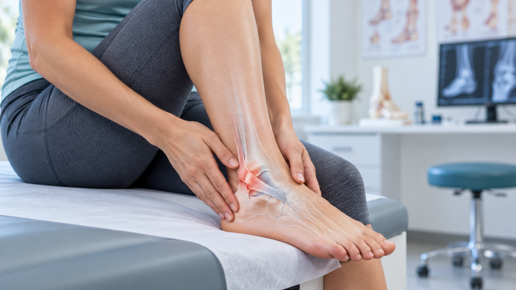
Ligaments connect bone to bone and help keep the ankle stable. The most common sprain follows an inversion injury, when the sole turns inward and the outer ankle ligaments are loaded. The anterior talofibular ligament is often involved, sometimes together with the calcaneofibular ligament. Less common patterns affect the inner ankle or the syndesmosis above the joint, often called a high ankle sprain.
Pain on the outside of the ankle is common, but location alone does not confirm the diagnosis. Bone tenderness, pain higher above the ankle, inability to bear weight or symptoms at the base of the foot can change the examination and the need for imaging.
When an Ankle Injury Needs Urgent Assessment
While minor sprains often improve with conservative rest and self-care, acute trauma can involve fractures, joint dislocations, or neurovascular compromise that require immediate medical evaluation.
-
Visible Deformity or Dislocation: The ankle joint appears misaligned, crooked, or sitting completely out of its natural position.
-
Neurovascular Changes: The foot feels unusually cold, looks pale or bluish, or presents with numbness, loss of sensation, or worsening pins-and-needles.
-
Open Fractures & Bleeding: Any open skin wound near the injury, continuous bleeding, or exposed bone fragments.
-
Severe, Rapid Swelling & Pain: Disproportionately intense pain that spikes immediately alongside sudden, extensive localized swelling.
-
Inability to Bear Weight: Complete inability to take more than a couple of steps or put any weight through the foot right after the incident.
-
Bony Point Tenderness: Sharp, localized tenderness directly over the malleoli (bony ankle bumps) or midfoot bones, indicating potential fracture patterns under Ottawa Ankle Rules.
-
Signs of Infection or Systemic Illness: Expanding redness, radiating heat, fever, or feeling unwell following an initial injury.
Hearing a “pop” does not definitively indicate a complete ligament tear, nor does the ability to limp a few paces entirely exclude a hairline fracture. A formal clinical examination and timely imaging are the most reliable ways to determine whether your injury requires immobilization, casting, or surgical stabilization.
Ankle Sprain Grades
Sprain grades describe ligament damage, but early swelling and pain can make self-grading unreliable. The table is a clinical guide, not a substitute for examination.
| Grade | Ligament change | Typical findings | Stability |
| Grade 1 | Stretching and microscopic tearing | Mild tenderness and swelling | Usually stable |
| Grade 2 | Partial ligament tear | Moderate pain, bruising and swelling | Some looseness may be present |
| Grade 3 | Complete ligament tear | Marked swelling, bruising and weight-bearing difficulty | Instability may be present |
How an Ankle Sprain Is Diagnosed
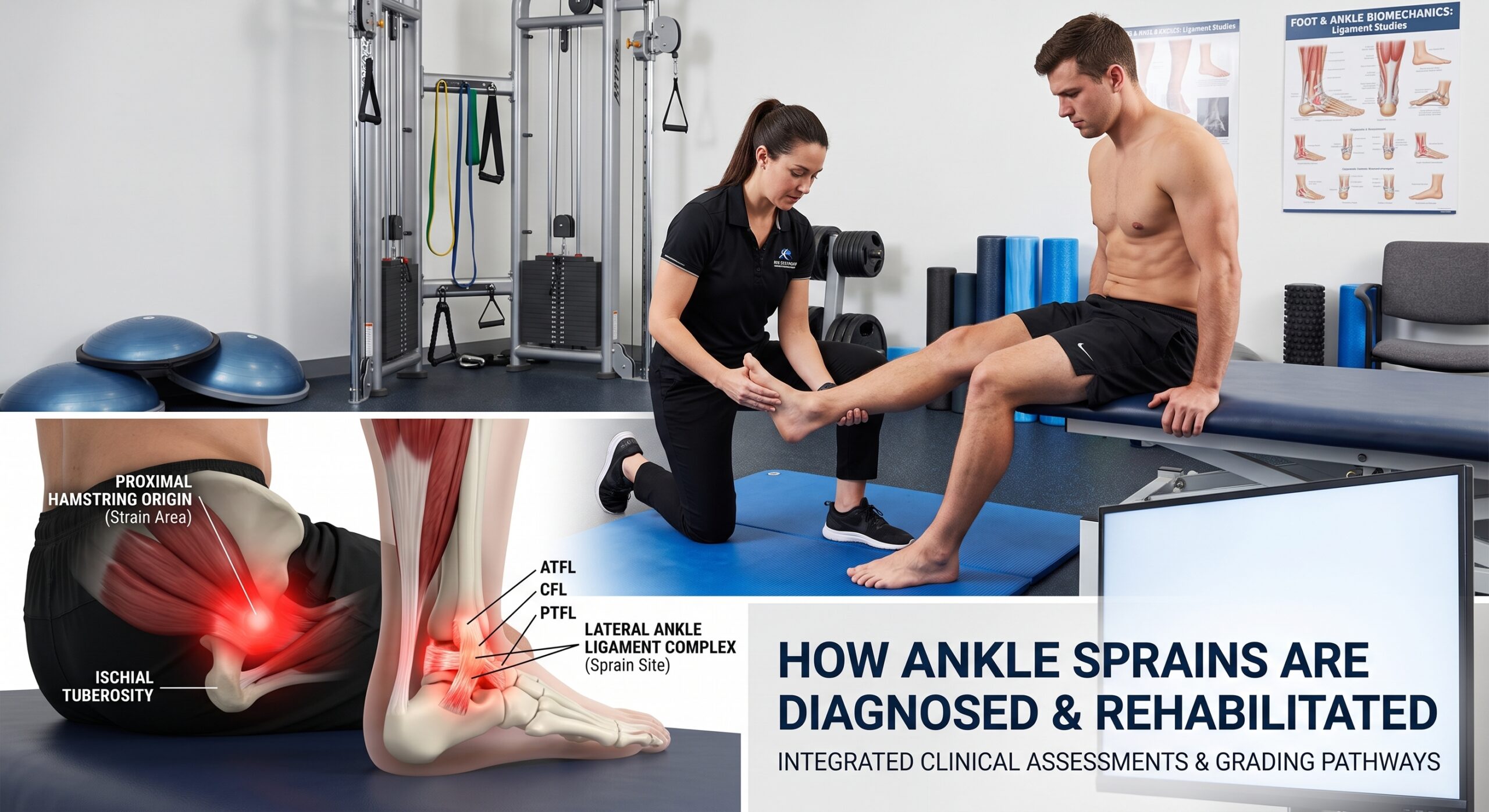
A comprehensive clinical assessment identifies which ligaments are involved, checks joint stability, and rules out complex trauma such as syndesmotic (“high ankle”) injuries, tendon ruptures, or occult fractures.
-
Injury Mechanism & History: The evaluation begins by establishing how the trauma occurred—whether the foot rolled inward (inversion) or outward (eversion), if direct contact was involved, where pain was immediately felt, and whether weight-bearing was possible after the event.
-
Timing & Severity Indicators: Assessing how quickly swelling, bruising, and mobility limitations developed helps distinguish a routine lateral ligament strain from bone fractures or syndesmotic compromise.
-
Physical & Stability Examination: The specialist examines both ankles side-by-side to compare contours, check capillary refill and circulation, pinpoint focal tenderness, and evaluate ligamentous laxity or joint play once pain allows safe stress testing.
-
Targeted Diagnostic Imaging: Routine imaging is not indicated for every minor twist, but advanced tools are deployed strategically:
-
X-ray: Ordered when clinical findings or Ottawa Ankle Rules suggest a potential fracture of the malleoli or metatarsals.
-
Ultrasound: Evaluates superficial ligament tears (such as the ATFL and CFL) as well as peroneal and Achilles tendon integrity dynamically.
-
MRI: Reserved for unresolved pain, suspected osteochondral cartilage lesions, tendon pathology, or high ankle sprains (syndesmotic tears) where detailed imaging dictates surgical versus non-surgical decisions.
-
-
Clinical Consensus: As emphasized by the American Academy of Orthopaedic Surgeons ankle sprain guide, diagnosis remains fundamentally clinical, with imaging reserved to exclude fractures or map extensive soft-tissue injury when symptoms warrant deeper investigation.
Early Ankle Sprain Treatment
Early management focuses on shielding damaged ligaments from additional strain, minimizing swelling, and maintaining gentle, safe mobility without enforcing prolonged, complete immobilization.
-
Joint Protection & Support: Safeguard the ankle against recurrent twisting using a semi-rigid brace, supportive athletic footwear, a walking boot, or crutches, tailored to the patient’s pain tolerance and joint stability.
-
Relative Rest & Load Management: Cease high-impact movements immediately—including running, jumping, and rapid directional changes—while preserving controlled daily movement within a comfortable, pain-free range.
-
Cryotherapy (Cold Application): Apply a towel-wrapped cold pack for brief intervals to numb acute pain, ensuring ice is never placed directly on bare skin or over areas with impaired sensation.
-
Comfortable Compression: Utilize a supportive elastic bandage to control localized swelling, ensuring it remains comfortable; loosen the wrap immediately if the foot develops numbness, pale discoloration, coolness, or heightened pain.
-
Limb Elevation: Elevate the injured ankle above heart level whenever resting to facilitate venous return and reduce acute interstitial fluid pooling.
-
Clinical Treatment Guidelines: These steps align with established NHS guidance on sprains and strains, which recommends phased protection, rest, ice, compression, and elevation (PRICE) during the initial acute phase, followed by progressive movement once pain allows.
-
Personalized Pain Relief: Always consult a doctor or pharmacist prior to using pain medications or non-steroidal anti-inflammatory drugs (NSAIDs), particularly for individuals with a history of gastrointestinal ulcers, renal disease, cardiovascular issues, pregnancy, anticoagulant therapies, or drug sensitivities.
Functional Rehabilitation
Rehabilitation should begin at the level the ankle can tolerate and progress according to pain, swelling, control and function. A generic exercise list cannot replace an assessment when the ankle is unstable or the diagnosis is uncertain.
Restore Movement
Gentle ankle movement can help limit stiffness once it is safe. Early exercises may include controlled ankle circles or tracing the alphabet with the foot. Movement should stay within a comfortable range and should not reproduce sharp pain.
Rebuild Strength
Calf, ankle and foot strength help control the joint during walking, landing and direction changes. Resistance is added gradually, often progressing from low-load isometric work to band exercises, calf raises and task-specific loading.
Retrain Balance and Position Sense
An ankle sprain can affect proprioception, the body’s awareness of joint position. Balance work may begin with supported single-leg standing and progress to unstable surfaces, reaching tasks and sport-specific reactions. This stage matters because pain relief alone does not restore control.
Prepare for Sport
Running, cutting, jumping and contact practice return in stages. Straight-line movement usually comes before rapid changes of direction. The progression should reproduce the demands of the athlete’s sport without causing new swelling, instability or a meaningful next-day flare.
Patients with a significant or recurrent sprain can review the clinic’s ligament injury assessment and treatment pathway for more information about diagnosis-led care.
Bracing Taping and Immobilization
A brace or tape may support the ankle during early walking and the return to higher-risk activity. The choice depends on the sprain pattern, swelling, footwear and sport. Support should not be so tight that it changes sensation or circulation.
More severe injuries may need a short period in a walking boot or another form of immobilization. Prolonged immobilization without a clear reason can contribute to stiffness and weakness, so protection should be reviewed and followed by an appropriate rehabilitation plan.
When Surgery Is Considered
Surgical intervention is rarely needed for an isolated, standard ankle sprain, but it becomes an important clinical option when mechanical instability persists or when associated joint structures are damaged.
-
Chronic Instability & Persistent Pain: Surgery may be indicated when chronic ankle instability (frequent giving way) and discomfort fail to improve after dedicated, progressive physical therapy and neuromuscular retraining.
-
Unstable Syndesmotic (High Ankle) Sprains: Severe syndesmosis disruption compromises the integrity of the lower leg bones (tibia and fibula), often requiring operative fixation or suture-button stabilization to restore joint mortise alignment.
-
Associated Structural Pathology: Operative care addresses concurrent intra-articular injuries, including displaced osteochondral cartilage lesions (talar dome defects), loose bodies, or peroneal tendon subluxation and tears.
-
Tailored Operative Plans: The specific procedure—ranging from minimally invasive arthroscopic debridement to anatomical ligament repair or reconstruction—is determined by the confirmed tissue pathology rather than a generic injury label.
-
Comprehensive Clinical Care: Learn more about our dedicated protocols by exploring Dr. Ahmed Azmy’s sports injury treatment service, outlining our specialized care pathway for complex ligament, tendon, and athletic injuries in Sharjah.
Recovery Time
Ankle sprain recovery varies widely based on injury severity, the specific ligaments involved, previous sprain history, rehabilitation consistency, and the physical demands of your daily routine or sport.
-
Mild (Grade I) Sprains: Typically show substantial improvement within one to three weeks with relative rest, swelling management, and early mobility exercises.
-
Moderate to Severe (Grade II & III) Sprains: Often require six to twelve weeks or longer before clearing an athlete for unrestricted training and cutting maneuvers.
-
Syndesmotic (High Ankle) Sprains: Involve the stabilizing ligaments between the tibia and fibula, necessitating a significantly slower, protected recovery pathway compared to standard lateral sprains.
-
Functional Milestone Tracking: Progression must be guided by objective functional milestones, such as pain-free single-leg hopping and full balance control, rather than rigid calendar dates.
-
When to Re-evaluate: Lingering joint swelling, sensations of the ankle giving way, sharp focal bone tenderness, or plateaued recovery indicate the need for clinical reassessment rather than forcefully pushing through high-intensity activities.

Return to Sport Criteria
A date alone does not show that an ankle is ready for sport. The decision should consider symptoms, movement, strength, balance, confidence and sport-specific performance.
| Area | Useful return marker |
| Symptoms | Minimal pain and no meaningful increase in swelling after loading |
| Movement | Near-normal ankle movement for the required activity |
| Strength | Good calf and ankle strength compared with the uninjured side |
| Balance | Controlled single-leg balance and landing mechanics |
| Sport tasks | Progressive running, jumping and direction changes completed with control |
| Response | No instability or significant flare later that day or the next morning |
How to Reduce the Risk of Another Sprain
Because a prior sprain remains the single greatest risk factor for future ankle trauma, completing a comprehensive conditioning plan is vital to restore mechanical stability and neuromuscular defense.
-
Complete Full-Phase Rehabilitation: Continue structured strength, calf capacity, and balance (proprioception) training even after daily walking becomes completely pain-free, ensuring the joint is prepared for unpredictable forces.
-
Graded Sport-Specific Loading: Reintroduce cutting, pivoting, decelerating, and jumping drills gradually rather than returning immediately to unrestricted competitive matches.
-
Surface-Appropriate Footwear: Select athletic shoes designed specifically for your playing surface (e.g., turf, court, or trail), and replace worn footwear promptly once midsole support, heel counter stiffness, or tread traction breaks down.
-
Dynamic, High-Speed Warm-Ups: Prepare the lower kinetic chain with active drills that mimic the speed, multidirectional agility, and demands of your sport, avoiding passive static stretching alone.
-
Prophylactic Bracing or Taping: Utilize supportive semi-rigid ankle braces or athletic taping during the initial return-to-sport phase or during high-risk pivot activities when advised by your specialist.
-
Prompt Clinical Reassessment: Arrange a follow-up consultation if your ankle repeatedly feels loose, gives way during routine maneuvers, or develops chronic, lingering post-exercise swelling.
-
Evidence-Based Prevention Guides: For comprehensive conditioning strategies, explore our practical clinical guide on how to prevent sports injuries and review the overview of common sports injuries.
When to Book an Orthopedic Assessment in Sharjah
Arrange an orthopedic assessment if you cannot walk comfortably, the ankle feels unstable, swelling is substantial, symptoms are not improving as expected, or the injury keeps recurring. Athletes should also seek guidance when a clear diagnosis and return-to-sport plan are needed for football, running, court sports or gym training.
Contact Dr Ahmed Azmy Clinic in Sharjah to arrange an ankle and sports injury assessment. Use urgent medical services instead of a routine appointment if there is deformity, loss of circulation or sensation, an open injury, severe trauma or rapidly worsening symptoms.
Medical Disclaimer
This article provides general education and does not diagnose an individual injury or replace care from a qualified healthcare professional. Treatment, medication, imaging and return-to-sport decisions must be individualized. Seek urgent medical care for severe trauma, deformity, an open wound, loss of circulation or sensation, or rapidly worsening symptoms.
Book an appointment now with Dr Ahmed Azmy in Sharjah.