Assess your pain
Dr. Ahmed Azmy
Fracture & Trauma Care · Sharjah

Upper Limb Fractures Treatment in Sharjah

Specialist diagnosis, fracture fixation and rehabilitation for shoulder, arm, elbow, forearm and wrist injuries.

Upper limb fractures can affect movement, work and daily activities. Dr. Ahmed Azmy provides specialist assessment, imaging review, conservative treatment, surgical fixation and structured rehabilitation in Sharjah.

Accurate Diagnosis Identify fracture pattern and associated injuries.
Stable Alignment Restore and maintain the correct bone position.
Nerve Protection Assess sensation, movement and hand circulation.
Functional Recovery Protect joint motion and support rehabilitation.
Dr. Ahmed Azmy orthopedic specialist in Sharjah
Orthopedic & Sports Injuries Specialist Dr. Ahmed Azmy Fracture and trauma care · Sharjah, UAE
Clinical Anatomy

What Are Upper Limb Fractures?

An upper limb fracture is a partial or complete break in one or more bones extending from the shoulder girdle to the wrist.

Treatment considers more than the fractured bone alone. Joint alignment, nearby ligaments, tendons, muscles, nerves, circulation, and overall arm function must also be assessed.
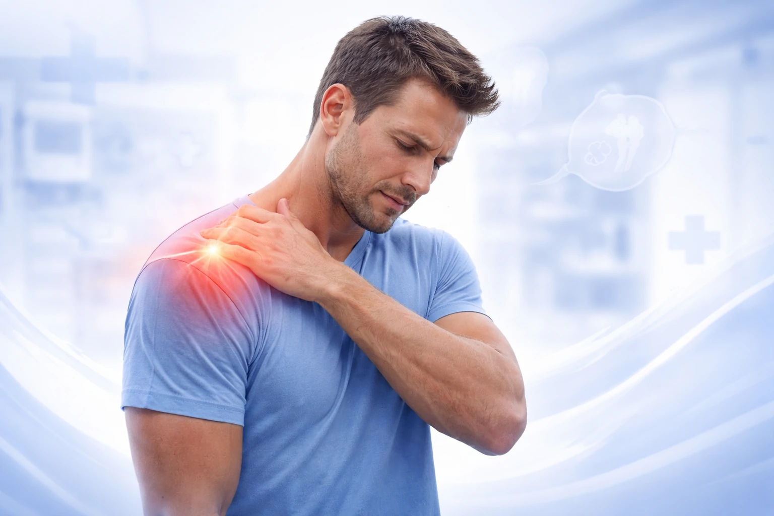
Patient with upper limb fracture injury
Upper Limb From shoulder
to wrist

Fractures may also affect nearby joints, soft tissues, nerves and blood vessels.

Upper limb fracture x-ray
01 Imaging Review
Clinical fracture assessment
02 Clinical Assessment
+
Clinical Perspective

A fracture is more than a broken bone

Specialists assess stability, displacement, joint involvement, soft tissue damage and neurovascular status before deciding treatment.

01 Stability

Can the fracture maintain normal alignment?

02 Joint Involvement

Does the fracture extend into a joint surface?

03 Neurovascular Status

Are nearby nerves and circulation protected?

Upper Limb Anatomy

One connected kinetic chain

Fracture assessment follows the limb from the shoulder girdle through the arm, elbow and forearm to the wrist.

01

Clavicle

The collarbone connecting the chest and shoulder.

02

Shoulder

Proximal humerus, scapula and surrounding joint structures.

03

Upper Arm

The humeral shaft extending between the shoulder and elbow.

04

Elbow

Distal humerus, radial head and olecranon region.

05

Forearm

The radius and ulna extending from elbow to wrist.

06

Wrist

Distal radius, distal ulna and the carpal bones.

!
Why early assessment matters

Prompt evaluation is particularly important when the arm appears deformed, the fingers feel numb or cold, significant swelling develops, or the joint may also be dislocated.

Fracture Patterns

Types of Upper Limb Fractures

Fractures are classified by the bone involved, the break location, alignment, stability and whether the injury extends into a joint.

Scroll or drag to explore
Clavicle fracture
01
Collarbone

Clavicle fracture

Often follows a fall onto the shoulder or outstretched hand. Pain, swelling and a visible bump may develop.

Proximal humerus fracture
02
Shoulder

Proximal humerus fracture

May cause severe shoulder pain, bruising and inability to raise the arm, commonly after a fall.

Scapular fracture
03
Shoulder Blade

Scapular fracture

Usually associated with high-energy impact and may occur with chest or other traumatic injuries.
Humeral shaft fracture
04
Upper Arm

Humeral shaft fracture

Can produce deformity and instability. The radial nerve runs close to this region and must be assessed carefully.
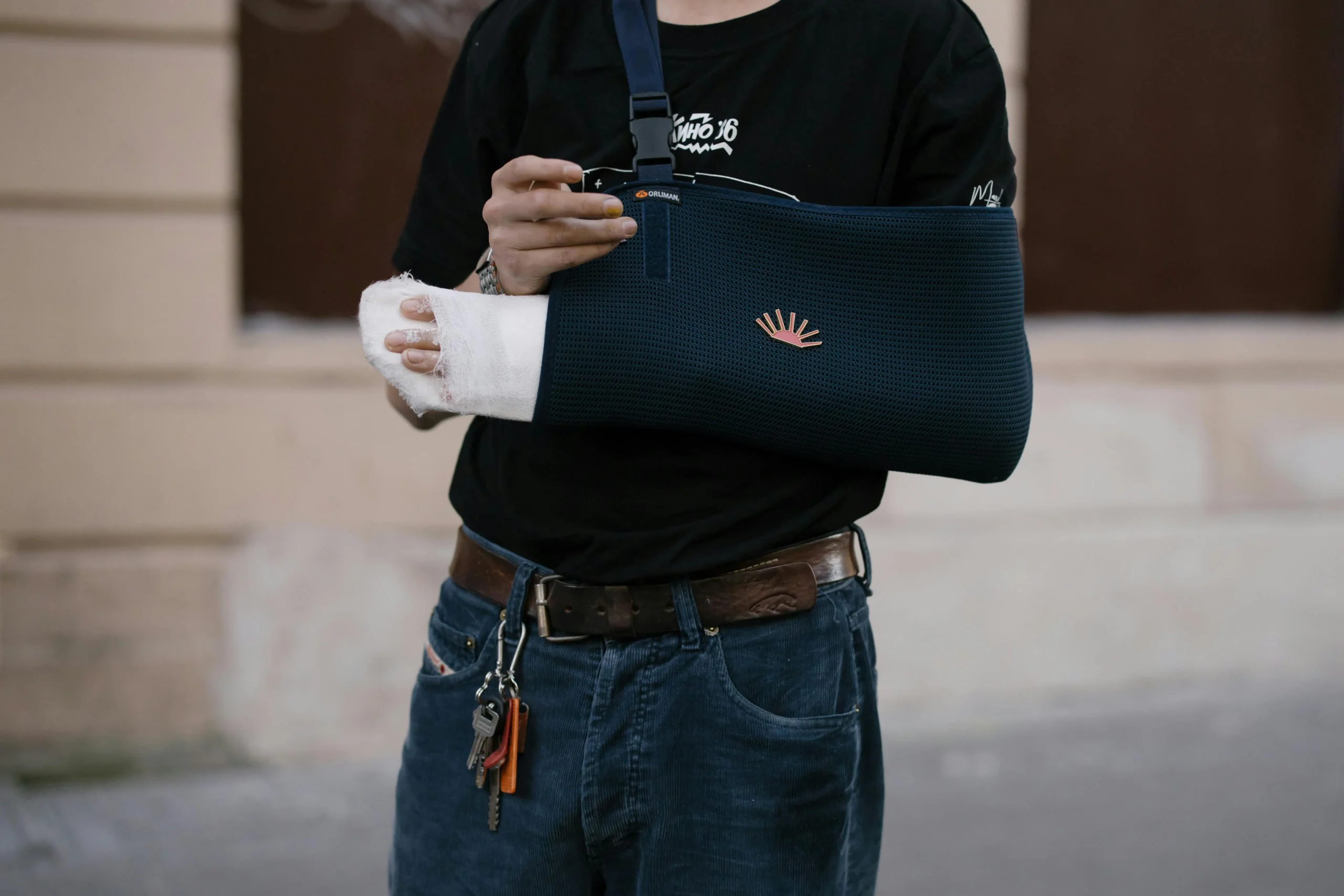
Distal humerus and olecranon fracture
05
Elbow

Distal humerus / Olecranon

Potentially complex joint fractures causing severe pain, restricted motion or inability to straighten the arm.
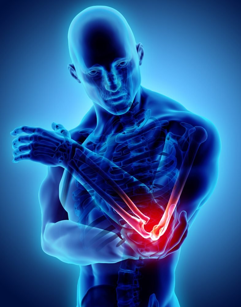
Radius and ulna fractures
06
Forearm

Radius & Ulna fractures

Correct length, alignment and rotation are essential for normal forearm movement.
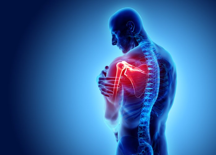
Distal radius and scaphoid fracture
07
Wrist

Distal radius / Scaphoid

May cause wrist deformity or subtle thumb-side pain. Some scaphoid fractures are not clear on initial X-rays.
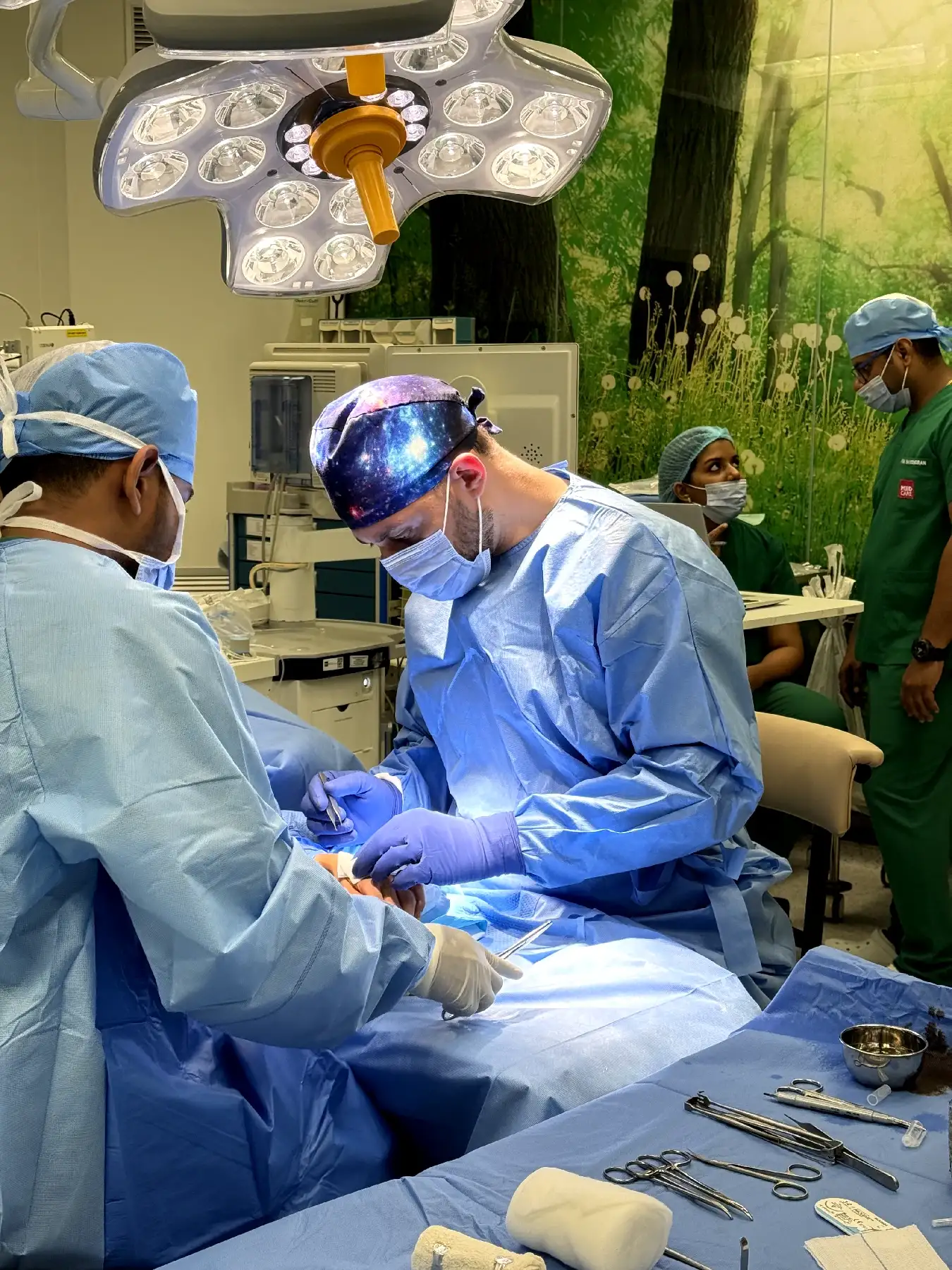
Open fracture and fracture-dislocation
08
Urgent Pattern

Fracture-dislocation / Open fracture

A broken bone combined with joint displacement or a skin wound may require urgent reduction and emergency treatment.

01
Shoulder Region

Shoulder Fractures

  • Clavicle fracture
  • Proximal humerus fracture
  • Scapular fracture
  • Shoulder fracture-dislocation
02
Elbow Region

Elbow Fractures

  • Distal humerus fracture
  • Radial head fracture
  • Olecranon fracture
  • Complex elbow fracture-dislocation
03
Forearm & Wrist

Forearm & Wrist

  • Radius and ulna fractures
  • Distal radius fracture
  • Distal ulna fracture
  • Scaphoid fracture
Recognise the Signs

Upper Limb Fracture Symptoms

Symptoms usually begin immediately after an accident, although subtle wrist injuries and stress fractures may become apparent more gradually.

Pain · Swelling · Deformity · Neurological Change
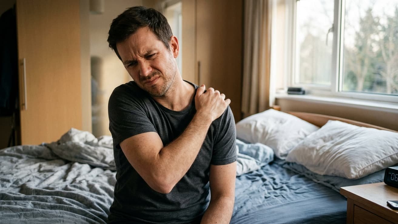
Patient with painful upper limb fracture
01 Primary Symptom Cluster
Most Common Presentation

Pain &
Loss of Function

Significant fractures commonly produce immediate pain accompanied by difficulty using the injured arm normally.

Clinical Signal Movement becomes limited
Quick Symptom Scan Pain-related limitations
01 Sudden severe pain
02 Pain with movement
03 Difficulty lifting the arm
04 Pain when gripping
05 Limited elbow movement
06 Reduced forearm rotation
02
Soft Tissue Response

Immediate Swelling

Inflammation may develop quickly after trauma and can increase pressure around the injured area.

  • Rapid swelling
  • Increasing tightness
  • Swollen fingers
  • Local tenderness
03
Tissue Trauma

Bruising

Bleeding beneath the skin can appear locally or spread away from the original injury over time.

  • Dark bruising
  • Bruising spreading down the arm
  • Discolouration near elbow or wrist
04
Alignment Change

Visible Deformity

Displacement or angulation may visibly alter the normal shape and alignment of the upper limb.

  • Abnormal angle
  • Shortened appearance
  • Prominent collarbone
  • Bent or displaced wrist
05
Neurovascular Warning

Nerve or Circulation Symptoms

Neurological or vascular symptoms require particular attention because they may indicate compression or injury.

  • Numbness or tingling
  • Weak wrist or finger movement
  • Pale or cold fingers
  • Reduced pulse
Urgent assessment may be required
Mechanism of Injury

Common Causes of Arm and Shoulder Fractures

Understanding how the injury happened helps predict associated damage and guides imaging and treatment.

01

Fall on an Outstretched Hand

Force travels through the wrist, forearm and elbow and may reach the shoulder.

  • Distal radius / scaphoid
  • Radial head / distal humerus
  • Proximal humerus / clavicle
02

Direct Impact

A direct blow can fracture the bone at the point of contact.

  • Shoulder or elbow fall
  • Sports equipment impact
  • Workplace / industrial trauma
03

Road Traffic Accident

High-energy trauma can produce displaced, open or multiple fractures.

  • Vehicle collision
  • Motorcycle accident
  • Crush injury
04

Sports Injury

Collision, awkward landing or forced joint rotation may cause fractures during high-speed and contact sports.

05

Fall from Height

A major fall can transmit substantial force through the upper limb, causing complex fractures or joint injuries.

06

Reduced Bone Density & Repetitive Loading

Osteoporosis or reduced bone strength can cause fractures after minor falls, while repeated loading may gradually create stress fractures.

FootballCyclingBasketballGymnasticsCombat SportsSkiing
Treatment Decision

Stable vs. Unstable Upper Limb Fractures

Fracture stability is one of the most important factors in treatment planning. It helps determine whether alignment can be safely maintained or mechanical stabilisation may be required.

Stability · Alignment · Joint Surface · Soft Tissues
Stable Fracture
Alignment Preserved

Alignment can be maintained

The fracture remains mechanically controlled and may heal successfully with appropriate protection, immobilisation and rehabilitation.

Typical pathway Conservative management
Unstable Fracture
Mechanical Instability

Stabilisation may be required

Fragments may shift, rotate or lose alignment, increasing the risk of joint damage, soft-tissue compromise and impaired healing.

Typical pathway Reduction or fixation
Stable Characteristics Clinical Factor Unstable Characteristics
01

Fragments remain in an acceptable position.

Bone Alignment
01

Displaced, angulated, shortened or rotated fragments.

02

Simple crack or minimally displaced break.

Fracture Pattern
02

Comminuted, segmental or significantly displaced pattern.

03

Joint surface remains congruent and well aligned.

Joint Involvement
03

Fracture extends into or disrupts the joint surface.

04

Skin and surrounding tissues remain intact.

Soft Tissues
04

Open wound, threatened skin or major soft-tissue injury.

05

Normal sensation, movement and blood flow.

Nerve & Circulation
05

Numbness, weakness or impaired circulation may be present.

06

Sling, cast, splint, brace and monitored rehabilitation.

Typical Management
06

Reduction, fixation or another stabilising procedure.

Precision Before Treatment

Upper Limb Fracture Diagnosis

A detailed diagnosis establishes fracture pattern, fragment position, joint involvement, stability and whether nerves, blood vessels or soft tissues have been damaged.

X-ray fracture imaging
X-Ray
CT scan fracture imaging
CT Scan
MRI fracture imaging
MRI
01

Patient History

  • Mechanism and timing
  • Fall, collision or direct impact
  • Arm position during injury
  • Previous fractures or surgery
  • Bone health and medication

Why it matters: predicts likely fracture pattern and associated injury.

02

Physical Examination

  • Swelling and bruising
  • Deformity, shortening or rotation
  • Open wounds or skin tenting
  • Associated dislocation

Identifies the injured region, fracture severity and urgency.

03

Nerve Assessment

  • Hand sensation
  • Wrist, finger and thumb movement
  • Grip and motor function
  • Numbness or weakness

Detects neurological compromise.

04

Circulation Assessment

  • Radial and ulnar pulses
  • Finger colour and temperature
  • Capillary refill
  • Swelling or circulation changes

Identifies impaired blood flow requiring emergency treatment.

05

Adjacent Joints & Soft Tissues

  • Shoulder and elbow stability
  • Forearm rotation
  • Tendon / ligament integrity
  • Joints above and below

Trauma may injure more than one region.

06

X-Ray Imaging

  • Fracture location and pattern
  • Displacement / angulation
  • Joint involvement
  • Associated dislocation

Primary investigation for most suspected upper-limb fractures.

07

CT Scan

  • Complex / comminuted fractures
  • Intra-articular fractures
  • 3D fragment position
  • Preoperative anatomy

Detailed information for complex injuries and surgical planning.

08

MRI

  • Occult fractures
  • Scaphoid fracture with unclear X-rays
  • Ligament, tendon or cartilage injury
  • Selected nerve / soft-tissue injury

Finds injuries that may not be visible on standard X-rays.

X-RayMost fractures, alignment, displacement and dislocation.
CT ScanComplex, comminuted and intra-articular fracture planning.
MRIOccult fractures and associated soft-tissue injury.
Emergency Assessment

When to Seek Immediate Medical Attention

Do not wait for a routine clinic appointment when severe deformity, an open wound, circulation changes, neurological symptoms or signs of compartment syndrome are present.

Urgent signs require prompt clinical assessment
Immediate Clinical Assessment

Some fracture symptoms should not wait.

Obtain emergency medical care if the injured arm is severely deformed, the skin is open, the hand becomes cold or pale, or neurological symptoms are worsening.

01 Open Injury Skin integrity compromised
02 Circulation Cold, pale or blue fingers
03 Neurology Numbness or weakness
04 Deformity Visible loss of alignment
01
Skin Integrity

Open Injury

Bone visible Contaminated wound Bleeding Threatened skin

Contamination increases infection risk and requires immediate treatment.

02
Alignment

Visible Deformity / Dislocation

Abnormal angle Shortening Joint displacement Skin tenting

Severe displacement may damage skin, nerves, blood vessels or joint surfaces.

03
Blood Flow

Circulation Problem

Pale fingers Cold hand Weak pulse Rapid swelling

Reduced blood flow requires immediate assessment to protect hand function.

04
Neurological Function

Nerve Problem

New numbness Loss of sensation Finger weakness Unable to move

New neurological changes may indicate nerve compression or direct nerve injury.

05
Tissue Pressure

Possible Compartment Syndrome

Escalating pain Tense forearm Pain moving fingers Numbness

Increasing tissue pressure can damage muscles and nerves if treatment is delayed.

06
High-Energy Trauma

Severe Trauma / Multiple Injuries

Road collision Fall from height Crush injury Multiple symptoms

High-energy trauma may require urgent hospital assessment for additional injuries.

Before Medical Assessment

Immediate First-Aid Guidance

Protect the injured arm and avoid manoeuvres that may worsen displacement or damage nearby tissues.

01
Keep the arm still

Leave it in the position in which it was found.

02
Provide support

Use a sling, towel or padded splint.

03
Remove restrictive items

Take off rings, watches and tight bracelets.

04
Protect open wounds

Cover them gently with a clean dressing.

05
Do not reposition bone

Never push exposed bone back through the skin.

06
Do not straighten deformity

Avoid attempting to realign the injured arm.

07
Use wrapped ice

Apply around the injured area without direct skin contact.

08
Prepare for possible surgery

Avoid food and drink if urgent surgery may be required, unless advised otherwise.

Restore Alignment · Protect Function

Treatment Options for Upper Limb Fractures

Treatment aims to restore alignment, achieve stable bone healing, protect nerves and blood vessels and preserve shoulder, elbow, wrist and hand function.

Fracture conservative care
01
Non-Surgical Protect the fracture while natural healing occurs
Stable / acceptably aligned

Stable, non-displaced or acceptably aligned fractures

Immobilisation Sling, shoulder immobiliser, cast, removable wrist brace or functional brace.
Closed Reduction Fragments may be realigned without an incision when appropriate.
Pain & Swelling Control Elevation, wrapped ice, finger movement and clinical pain relief.
Follow-Up Imaging Repeat X-rays confirm that alignment remains acceptable.
Controlled Early Movement Movement begins when clinically safe to reduce stiffness.
Rehabilitation Progressive movement, grip strengthening, resistance and return-to-function work.
Advanced fixation

Unstable, displaced, open or joint-involving injuries

Open Reduction & Internal Fixation Plates, screws or other implants restore and maintain alignment.
Percutaneous Fixation Pins, wires or screws through smaller incisions under imaging guidance.
Intramedullary Fixation A rod or nail may be used for selected humeral or forearm patterns.
External Fixation Useful for open fractures, severe swelling or major soft-tissue damage.
Joint Reconstruction Restores joint-surface alignment, stability and movement.
Joint Replacement May be considered for selected severe shoulder or elbow fractures.
Functional Recovery

Recovery Timeline and Functional Phases

Recovery varies according to the bone, fracture pattern, age, bone quality, treatment method, soft-tissue injury and rehabilitation needs.

24–72h

Emergency protection

Immobilisation, imaging, pain control and neurovascular checks.

W1–2

Early stabilisation

Cast, brace or wound review and repeat X-rays where needed.

W2–6

Early bone healing

Maintain alignment and gradually reduce swelling.

W6–8

Healing review

Assess tenderness, alignment and healing before progression.

W8–12

Mobility restoration

Progressive joint movement and light strengthening.

M3–4

Functional recovery

Grip work, resistance exercise and occupational rehabilitation.

M4–6

Advanced rehab

Work and sport-specific progression.

M6–12

Complex recovery

Continued improvement for complex joint or nerve injuries.

Phase 1

Protection & Pain Control

  • Immobilisation
  • Swelling reduction
  • Skin, nerve and circulation monitoring
Phase 2

Mobility Restoration

  • Controlled joint movement
  • Scar and swelling management
  • Prevent avoidable stiffness
Phase 3

Strength Recovery

  • Grip strengthening
  • Progressive resistance
  • Joint control and endurance
Phase 4

Return to Function

  • Work simulation
  • Sports progression
  • Lifting and endurance training
Injury Factors
  • Open / high-energy injury
  • Displacement / comminution
  • Joint involvement
  • Nerve or vascular injury
Medical Factors
  • Reduced bone density
  • Smoking / nicotine
  • Diabetes
  • Nutrition or previous stiffness
Recovery Factors
  • Missed follow-up
  • Premature loading
  • Poor rehabilitation adherence
  • Infection or wound issues
Why Follow-Up Matters

Complications of Untreated or Malaligned Fractures

A fracture that is not diagnosed, reduced or monitored appropriately can affect the entire upper limb.

01

Malunion

  • Abnormal bone position
  • Visible deformity
  • Reduced rotation
  • Weak grip
  • Persistent pain

May change how force passes through the shoulder, elbow, forearm or wrist.

02

Delayed Union or Nonunion

  • Persistent pain
  • Slow or failed healing
  • Possible implant failure
  • Revision fixation or bone grafting
03

Joint Stiffness & Contracture

  • Reduced shoulder / elbow motion
  • Loss of forearm rotation
  • Difficulty dressing or working
  • Delayed return to sport
04

Post-Traumatic Arthritis

  • Chronic pain and swelling
  • Grinding or catching
  • Movement restriction
  • Cartilage degeneration
05

Nerve Injury

  • Persistent numbness
  • Neuropathic pain
  • Weakness or wrist drop
  • Loss of dexterity
06

Infection

  • Higher risk after open fracture
  • Wound redness / discharge
  • Delayed healing
  • Possible bone infection
07

Circulation Problems

  • Arterial compression / damage
  • Threat to muscle, nerve or skin
  • May require urgent restoration of flow
08

Compartment Syndrome

  • Escalating pain
  • Tense swelling
  • Pain with passive finger movement
  • Increasing weakness
09

Complex Regional Pain Syndrome

  • Persistent disproportionate pain
  • Swelling
  • Temperature / colour change
  • Stiffness and sensitivity
10

Tendon Irritation or Rupture

  • Pain during tendon movement
  • Clicking / catching
  • Reduced finger, thumb or shoulder movement
11

Avascular Necrosis

  • Bone collapse
  • Persistent pain
  • Progressive stiffness
  • Possible further surgery
Specialist Fracture Care

Why Choose Dr. Ahmed Azmy in Sharjah?

Upper limb trauma demands precise decision-making. The objective is not simply fracture union, but restoration of alignment, protection of nerves and joints, and practical use of the entire arm.

Dr. Ahmed Azmy
Dr. Ahmed Azmy

Orthopedic & Sports Injuries Specialist

Specialist fracture assessment for patients from Sharjah, Ajman and across the UAE.

15+Years of orthopedic and sports injury experience
40K+Patients treated
2,500+Advanced orthopedic procedures performed
Fellowship-trained in GermanyExperience in arthroscopy, fracture surgery and joint reconstruction.

A Patient-Centred Fracture Pathway

Accurate EvaluationMechanism, fracture pattern, soft tissues, neurological status and functional priorities.
Clear Treatment ExplanationStability, options, recovery and warning signs explained directly.
Individualised Surgical PlanningFixation technique selected for the fracture pattern and patient needs.
Functional RehabilitationProtect healing while reducing stiffness, weakness and loss of confidence.
Patient Success Stories

What Our Patients Say About Dr. Ahmed Azmy

Real experiences from patients who underwent ACL reconstruction and advanced knee treatments in Sharjah, restoring their lifestyle, mobility, and confidence.

★★★★★

"Dr. Ahmed Azmy is the best ACL surgeon in Sharjah. I returned to football just 7 months after my arthroscopic reconstruction surgery. Incredible care!"

- Tareq Al-Shamsi
★★★★★

"Highly professional doctor. He diagnosed my partial ACL tear accurately using MRI and saved me from an unnecessary operation with great physiotherapy."

- Sarah Michael
★★★★★

"After my sports injury, my knee was giving way completely. Dr. Azmy performed an excellent repair. Stability is now 100% perfect."

- Firas El-Din
★★★★★

"Excellent experience at the clinic in Sharjah. The care protocol before and after my ACL reconstruction surgery was structured and highly reassuring."

- Amna Al-Mansoori
★★★★★

"Dr. Ahmed is very patient and knowledgeable. He explained my graft options clearly and built a personalized rehab plan for my athletic goals."

- John Fletcher
★★★★★

"I traveled from Ajman to see Dr. Azmy for my chronic knee instability. His advanced minimally invasive approach made my recovery time very fast."

- Khalid Omar
★★★★★

"The gold standard of knee care in the UAE. His team is extremely professional and the post-operative follow-up is exceptionally detailed."

- Mustafa Reda
★★★★★

"Incredible results with PRP therapy combined with focused neuromuscular training. My knee swelling disappeared entirely in a few weeks."

- Elena Rostova
★★★★★

"Dr. Ahmed Azmy is the best ACL surgeon in Sharjah. I returned to football just 7 months after my arthroscopic reconstruction surgery. Incredible care!"

- Tareq Al-Shamsi
★★★★★

"Highly professional doctor. He diagnosed my partial ACL tear accurately using MRI and saved me from an unnecessary operation with great physiotherapy."

- Sarah Michael
★★★★★

"After my sports injury, my knee was giving way completely. Dr. Azmy performed an excellent repair. Stability is now 100% perfect."

- Firas El-Din
★★★★★

"Excellent experience at the clinic in Sharjah. The care protocol before and after my ACL reconstruction surgery was structured and highly reassuring."

- Amna Al-Mansoori
★★★★★

"Dr. Ahmed is very patient and knowledgeable. He explained my graft options clearly and built a personalized rehab plan for my athletic goals."

- John Fletcher
★★★★★

"I traveled from Ajman to see Dr. Azmy for my chronic knee instability. His advanced minimally invasive approach made my recovery time very fast."

- Khalid Omar
★★★★★

"The gold standard of knee care in the UAE. His team is extremely professional and the post-operative follow-up is exceptionally detailed."

- Mustafa Reda
★★★★★

"Incredible results with PRP therapy combined with focused neuromuscular training. My knee swelling disappeared entirely in a few weeks."

- Elena Rostova
★★★★★

"Outstanding orthopedic surgeon. My complete ACL tear plus meniscus damage was treated with high precision via arthroscopy. Highly recommended!"

- Saeed Al-Hajri
★★★★★

"Dr. Azmy gave me my life back. The intense pain when walking and knee catching sensations are completely gone after his treatment protocol."

- Maria Santos
★★★★★

"Brilliant experience. Functional testing and strict rehabilitation milestones guided by Dr. Ahmed helped me return to basketball safely."

- Youssef Karim
★★★★★

"A master of knee preservation. His conservative management plan for my acute strain was highly effective. No pain, full motion restored."

- Fatima Al-Ali
★★★★★

"If you want accurate diagnosis and a clear recovery roadmap for knee instability, Dr. Ahmed Azmy is absolutely the right choice."

- Arthur Pendelton
★★★★★

"The arthroscopic surgery was seamless. Minimal pain post-op, and my range of motion is improving faster than expected in physical therapy."

- Naser Hamad
★★★★★

"Very trustworthy specialist. He won't recommend surgery unless it is strictly necessary. His ethical approach is highly appreciated."

- Dr. Robert Cole
★★★★★

"Dr. Ahmed's expertise in treating high-impact sports injuries gave me the confidence to push through my ACL recovery phases safely."

- Ziad El-Mallah
★★★★★

"Outstanding orthopedic surgeon. My complete ACL tear plus meniscus damage was treated with high precision via arthroscopy. Highly recommended!"

- Saeed Al-Hajri
★★★★★

"Dr. Azmy gave me my life back. The intense pain when walking and knee catching sensations are completely gone after his treatment protocol."

- Maria Santos
★★★★★

"Brilliant experience. Functional testing and strict rehabilitation milestones guided by Dr. Ahmed helped me return to basketball safely."

- Youssef Karim
★★★★★

"A master of knee preservation. His conservative management plan for my acute strain was highly effective. No pain, full motion restored."

- Fatima Al-Ali
★★★★★

"If you want accurate diagnosis and a clear recovery roadmap for knee instability, Dr. Ahmed Azmy is absolutely the right choice."

- Arthur Pendelton
★★★★★

"The arthroscopic surgery was seamless. Minimal pain post-op, and my range of motion is improving faster than expected in physical therapy."

- Naser Hamad
★★★★★

"Very trustworthy specialist. He won't recommend surgery unless it is strictly necessary. His ethical approach is highly appreciated."

- Dr. Robert Cole
★★★★★

"Dr. Ahmed's expertise in treating high-impact sports injuries gave me the confidence to push through my ACL recovery phases safely."

- Ziad El-Mallah

FAQ

Not every fracture requires an operation. Surgery is more likely when the fracture is displaced, unstable, open, comminuted, extends into a joint or is associated with a dislocation. It may also be recommended when the skin is threatened, circulation or nerve function is affected, or acceptable alignment cannot be maintained in a cast. The final recommendation is based on the specific bone, fracture pattern, patient age, bone quality and functional requirements.

All three injuries can cause pain, swelling and loss of movement, so symptoms alone may not provide a reliable diagnosis. A fracture may produce focal bone tenderness, deformity, bruising or inability to use the arm. A sprain affects ligaments, while a dislocation occurs when the joint surfaces lose their normal alignment. Upper limb fractures and dislocations can occur at the same time, so a deformed joint should be immobilised and assessed urgently.

X-rays confirm most upper limb fractures and show alignment, displacement and joint involvement. CT may be required for complex, comminuted or intra-articular fractures and for surgical planning. MRI may be used when a scaphoid or other occult fracture is suspected despite unclear X-rays, or when ligament, tendon, cartilage or soft-tissue injury must be assessed.

The duration varies according to the bone, fracture stability, patient age, treatment method, bone quality and follow-up imaging. Removing protection too early can allow displacement, while keeping a joint immobilised longer than necessary can increase stiffness. The support should be adjusted or removed only when the fracture is clinically stable and the treating specialist confirms that movement can safely increase.

Rehabilitation begins at different times for different fractures. Finger movement may start early to reduce swelling, while shoulder, elbow, forearm or wrist exercises are introduced according to fracture stability and fixation security. Starting too aggressively can disturb the fracture; delaying permitted movement can contribute to stiffness. The plan should specify permitted motion, lifting restrictions and progression to strengthening.

Recovery depends on the type and severity of the nerve injury. A compressed, stretched or bruised nerve may gradually recover over weeks or months. A severely damaged or divided nerve may require nerve-conduction studies, electromyography, imaging or surgical exploration. New or worsening numbness, weakness, cold fingers or colour change should be assessed urgently rather than observed at home.

Do not drive while the arm is immobilised, medication affects alertness or you cannot control the vehicle safely. Return to work depends on whether the role is office-based or physically demanding. Keep non-waterproof casts and dressings dry, and follow wound-care instructions after surgery. Elevating and supporting the arm with pillows can improve sleep during the early stage. Clearance should be based on the specific fracture rather than a fixed number of days.

Return requires more than the absence of pain. The fracture should show adequate healing, and the patient should demonstrate functional range of motion, strength, grip, joint stability and safe sport-specific movement. Non-contact conditioning may begin earlier than collision sports or heavy lifting. Returning before sufficient healing increases the risk of displacement, refracture, implant failure or another injury caused by weakness and poor control.

Regain Arm Function

Regain Full Arm Functionality and Strength

The earlier the injury is accurately classified, the sooner the right plan can begin.

01

Restore Alignment

Re-establish the correct position of bone and joint.

02

Protect Nerves & Circulation

Identify and manage neurological or vascular compromise.

03

Select the Right Treatment

Choose conservative care or fixation according to stability.

04

Rebuild Function

Restore movement, strength, grip and practical arm use.

Injured Your Shoulder, Arm, Elbow, Forearm or Wrist?

Get expert evaluation and a personalised fracture treatment plan.

Restore alignment. Protect function. Recover with confidence with an Upper Limb Fracture Specialist in Sharjah.

Contact & Location

See Dr. Ahmed Azmy in Sharjah

Specialist orthopedic assessment at Medcare Hospital Sharjah, with direct access to clinical evaluation, imaging and personalised treatment planning.

Medcare Hospital Sharjah · Bu Danq
Clinic Location Medcare Hospital Sharjah Sheikh Saqr bin Al Qasimi · Bu Danq · Qasimia · Sharjah
Orthopedic consultation in Sharjah
01
Specialty Orthopedics & Sports Injuries
02
Location Sharjah, UAE
03
Hospital Medcare Hospital Sharjah
04
Appointment Online & Direct Booking
Dr. Ahmed Azmy Orthopedic & Sports Injuries logo.

Redefining Orthopedic Excellence in the UAE. Dr. Ahmed Azmy known as The Best Ortho Surgeon in Sharjah offers world-class, compassionate, and personalized treatments for sports injuries and advanced joint reconstruction.

Contact Us

Powered By ICONCLY Medical © 2026 . All rights reserved.

Patient Connection Hub