Left Shoulder Pain Causes Warning Signs and Treatment
Left shoulder pain is commonly linked to the rotator cuff, bursa, joint, biceps tendon or neck. The safest next step depends on...
Left shoulder pain is commonly linked to the rotator cuff, bursa, joint, biceps tendon or neck. The safest next step depends on how the pain began, which movements reproduce it and whether it is accompanied by weakness, numbness, breathing difficulty or chest discomfort.
Left shoulder pain may begin in the shoulder joint or surrounding tendons, muscles and bursae. It can also originate in the neck or, less commonly, be referred from another area of the body. The location alone cannot identify the cause. How the pain started, which movements reproduce it, and whether it is accompanied by weakness, stiffness, numbness or chest symptoms are more useful clues.
This guide explains common causes, warning signs, diagnostic steps and treatment options. It is educational and does not replace a medical examination. If shoulder discomfort occurs with possible symptoms of a heart attack or another emergency, seek emergency medical care rather than waiting for an orthopedic appointment.
What Does Pain in the Left Shoulder Mean
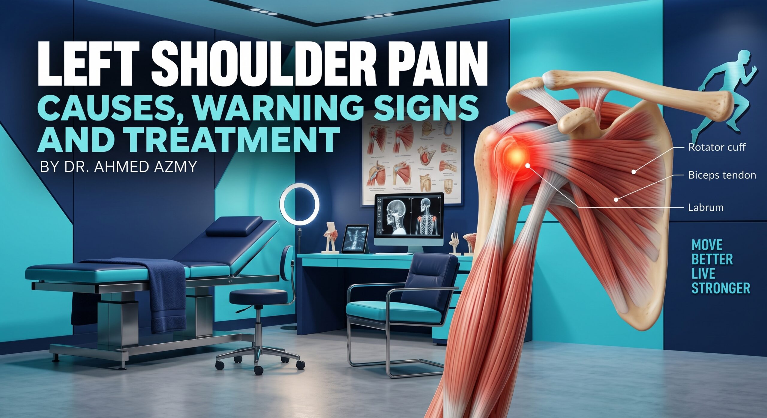
The shoulder is a highly mobile system formed by bones, joints, the rotator cuff, biceps tendon, joint capsule, labrum, bursae and the muscles that control the shoulder blade. Pain may be felt over the front or side of the shoulder, on top of the joint, around the shoulder blade or down the upper arm. Nerves from the cervical spine can also create pain, tingling, numbness or weakness in the shoulder and arm.
The fact that the pain is on the left side does not automatically make it heart-related, and it does not automatically prove that the shoulder itself is the source. A careful assessment considers the complete symptom pattern.
Common Orthopedic Causes of Left Shoulder Pain
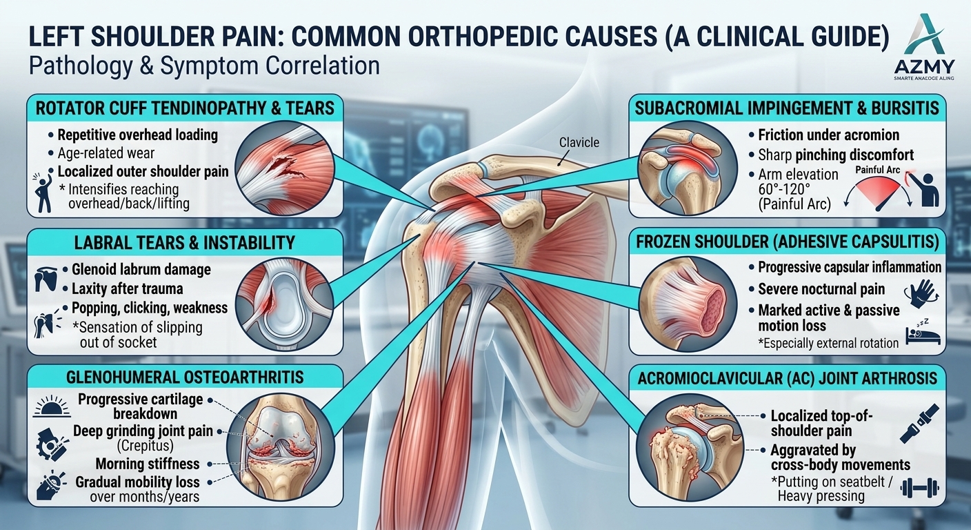
Shoulder pain arising directly from the glenohumeral joint or surrounding soft tissues typically results from tendon degeneration, mechanical impingement, capsular tightening, or structural trauma:
Rotator cuff tendinopathy and shoulder impingement
The rotator cuff is a group of tendons that helps raise and stabilize the arm. Irritation in these tendons or the tissues in the subacromial space may cause pain when reaching overhead, lifting an object or lowering the arm. The pain is often felt at the front or side of the shoulder and may be worse at night or when lying on the affected side.
Rotator cuff tear
A rotator cuff tear may develop after a fall or sudden force, or gradually as the tendon changes over time. Pain can be accompanied by true weakness, difficulty raising the arm and reduced function. Not every tear requires surgery, but sudden weakness after an injury should be assessed promptly. Learn more about the clinic’s rotator cuff tear assessment and treatment.
Frozen shoulder
Frozen shoulder (adhesive capsulitis) typically causes progressive deep aching followed by marked global stiffness. Both active movement and passive movement (assisted by an examiner) become mechanically restricted. Daily tasks such as dressing, reaching behind the back, and lifting the arm overhead become increasingly difficult. The condition often progresses through distinct stages (freezing, frozen, and thawing) and requires a stage-matched management plan based on pain severity and capsular restriction. Read more about frozen shoulder care.
Distinguishing Rotator Cuff Tears from Frozen Shoulder
Because both conditions limit elevation and cause severe discomfort, distinguishing tendon damage from capsular contracture relies on assessing active versus passive mobility:
| Clinical Feature | Rotator Cuff Tear | Frozen Shoulder (Adhesive Capsulitis) |
| Underlying Pathology | Structural fraying, degeneration, or rupture of rotator cuff tendon fibers. | Thickening, inflammation, and contracture of the glenohumeral capsule. |
| Active Range of Motion | Severely limited or weak; the patient cannot lift the arm unassisted. | Mechanically blocked; arm stops abruptly due to tight capsular constraint. |
| Passive Range of Motion | Preserved or near-normal; the arm moves freely when lifted by an examiner. | Severely restricted; the joint remains physically locked even with assistance. |
| Earliest Restricted Movement | Overhead abduction and resisted forward flexion. | External rotation (turning the forearm outward away from the waist). |
| Night Pain Pattern | Pain flares up primarily when direct pressure is placed on the affected side. | Deep, unrelenting throbbing that regularly wakes the patient regardless of position. |
| Primary First-Line Strategy | Rotator cuff reload exercises and periscapular stabilization. | Capsular preservation, anti-inflammatory intervention, and hydrodilatation/corticosteroid care. |
Biceps tendon irritation
The long head of the biceps tendon passes through the front of the shoulder. Irritation may produce pain in the front of the joint, especially during pulling, carrying, lifting or repeated overhead activity. It may occur alone or together with a rotator cuff problem.
Shoulder instability or dislocation
A previous dislocation or ligament injury can leave the shoulder feeling loose, unreliable or as if it may slip out of place. Some people notice painful clicking or apprehension in particular arm positions. A visibly deformed shoulder after trauma is an urgent problem and should not be pushed back into place without medical care. The clinic also provides information about dislocated shoulder treatment.
Arthritis and acromioclavicular joint pain
Arthritis may cause a deep ache, stiffness, and reduced movement. Pain directly on top of the shoulder, where the collarbone meets the shoulder blade, can arise from the acromioclavicular (AC) joint. Symptoms may follow an old injury or become more noticeable with loading across the joint.
Because treatments and mechanical triggers differ significantly between joint surfaces, a clinical evaluation distinguishes superficial AC joint wear from deep ball-and-socket degeneration:
| Diagnostic Feature | AC Joint Arthrosis | Glenohumeral Osteoarthritis |
| Exact Anatomical Location | Top of the shoulder where the collarbone (clavicle) joins the shoulder blade (acromion). | Deep inside the primary ball-and-socket joint (humeral head and glenoid cavity). |
| Pain Characteristics | Sharp, pinpoint tenderness directly over the bony prominence on top of the shoulder. | Diffuse, deep, dull joint ache felt centrally within the shoulder complex. |
| Provocative Movements | Cross-body reach (e.g., pulling a seatbelt, reaching across the chest) and overhead pressing. | Multi-directional movement, arm rotation, reaching behind the back, and lifting. |
| Mechanical Signs | Localized bony enlargement, tenderness to direct pressure, and pain during cross-body adduction. | Pronounced morning stiffness, audible crunching or grinding (crepitus), and global loss of range of motion. |
| First-Line Clinical Management | Cross-body load modification, targeted periscapular control, and localized ultrasound-guided AC joint injections. | Rotator cuff conditioning, joint-glide preservation, anti-inflammatory or regenerative injections, and joint replacement in advanced wear. |
Can Shoulder Pain Come From the Neck or Another Area?
Pain felt in the left shoulder does not always originate within the shoulder joint itself; irritation along the cervical spine or internal thoracic structures frequently refers pain down the shoulder blade and arm:
-
Cervical Radiculopathy (Pinched Neck Nerve): Compression of the C5, C6, or C7 nerve roots produces burning, shooting pain that radiates across the trapezius down past the elbow, often accompanied by numbness, tingling in the fingers, or thumb weakness.
-
Neck-Movement Provocation: If rotating, tilting, or extending your neck reproduces or worsens the shoulder symptoms (positive Spurling sign), the primary cause is cervical rather than shoulder joint pathology.
-
Diaphragmatic & Abdominal Referral: Irritation of the phrenic nerve from subdiaphragmatic issues (such as splenic injury, gastric conditions, or post-laparoscopic gas retention) can cause sharp referred pain to the top of the left shoulder (Kehr’s sign).
-
Myofascial Trigger Points: Chronic muscular tension in the levator scapulae, upper trapezius, and rhomboids commonly mimics deeper joint pain due to prolonged postural strain and desk work.
When Shoulder Pain May Be an Emergency
Because nerve pathways from the heart and the left shoulder share overlapping segments in the spinal cord, pain felt in the left shoulder or upper arm can be a classic warning sign of a life-threatening cardiovascular event rather than an orthopedic injury.
-
Acute Cardiac Signs (Heart Attack / Angina): Call emergency services immediately if left shoulder or arm ache occurs alongside crushing chest pressure, shortness of breath, sudden dizziness, profuse cold sweats, nausea, or pain radiating into the neck, jaw, or back.
-
Non-Mechanical Pain Pattern: Discomfort that begins without physical exertion or direct trauma, does not change when moving the shoulder joint, and feels like deep pressure or heaviness demands urgent emergency evaluation rather than an orthopedic appointment.
-
Traumatic Joint Deformities: Rapid medical assessment is also needed if a high-energy fall or collision causes an obvious visible dislocation, abnormal step-off at the collarbone, or total inability to move the arm.
-
Acute Vascular & Nerve Warning Signs: A hand or forearm that becomes cold, pale, bluish, or loses pulse signals arterial compromise, while sudden progressive numbness or hand drop requires urgent neurological review.
Symptoms That Help Identify the Cause

A specialist looks for patterns rather than relying on the word pain. Before an appointment, note when the problem began, whether there was an injury, what movements reproduce it and how it affects sleep, dressing, driving, work or sport.
- Pain during a particular arc of lifting may occur with rotator cuff or subacromial irritation.
- True weakness, rather than movement limited only by pain, may indicate a significant tendon or nerve problem.
- Loss of active and passive movement supports a stiffness-dominant condition such as frozen shoulder or arthritis.
- Clicking, slipping or apprehension may point toward instability or another mechanical problem.
- Tingling, numbness, burning pain or symptoms into the hand may indicate nerve involvement.
- Pain that is unrelated to shoulder movement requires consideration of referred or non-orthopedic causes.
How Shoulder Pain Is Diagnosed

An accurate diagnosis begins with a systematic clinical evaluation to isolate the damaged structure, determine joint biomechanics, and rule out referred symptoms from the neck or thoracic spine before selecting targeted imaging.
-
Comprehensive Physical Examination: The specialist assesses localized point tenderness, compares active versus passive range of motion, tests rotator cuff muscle strength, and performs targeted provocative tests (such as Hawkins-Kennedy or Neer impingement maneuvers) to reproduce symptoms safely.
-
Cervical Spine & Neurological Screening: Reflexes, sensory dermatomes, and neck motion (including Spurling’s test) are checked to confirm whether the pain originates locally within the shoulder joint or is referred from a pinched cervical nerve root.
-
First-Line Plain X-Rays: Routine weight-bearing and specialized radiographic views evaluate bone alignment, detect acromial bone spurs, identify calcific deposits, and assess joint space narrowing from glenohumeral or AC joint arthritis.
-
Dynamic High-Resolution Ultrasound: A non-invasive, cost-effective tool that assesses the subacromial bursa and rotator cuff tendons in real time as the arm moves, identifying dynamic impingement, tendon swelling, and fluid accumulation.
-
Targeted Magnetic Resonance Imaging (MRI): Advanced imaging is reserved for complex presentations, suspected labral tears (SLAP lesions), surgical planning for retracted rotator cuff tears, or when conservative care does not yield expected progress after several weeks.
-
Clinical Correlation Over Scan Reports: Not every patient requires an immediate MRI, as asymptomatic tears and age-related changes are common on scans after age 40; imaging findings are always interpreted alongside functional loss and physical examination results.
Treatment Options
Treatment depends on the diagnosis, severity, loss of function, activity requirements and whether the condition is traumatic, degenerative or referred. The aim is not only to reduce pain but also to restore safe movement and strength.
Activity modification and pain control
A short-term reduction in the movement that repeatedly aggravates symptoms can calm an irritable shoulder while avoiding prolonged immobilization. A clinician or pharmacist can advise whether a pain reliever or anti-inflammatory medicine is appropriate, as suitability varies with medical conditions and other medicines.
Physiotherapy and rehabilitation

Rehabilitation may include mobility work, rotator cuff and shoulder-blade strengthening, posture or movement changes and graded exposure to work, gym or sport demands. Exercises should match the diagnosis and current irritability rather than being copied from a generic strenuous program.
Injection treatment
A targeted injection may be considered for selected painful or inflammatory conditions when the diagnosis and expected benefit are clear. It is not a universal treatment for all shoulder pain and should form part of a broader plan when appropriate.
Surgery
Arthroscopic or open surgery may be considered for selected traumatic injuries, significant tears, recurrent instability or symptoms that continue despite appropriate non-surgical care. The decision is based on the structural problem, function, goals and response to treatment rather than pain alone. Explore the clinic’s shoulder arthroscopy services and non-surgical orthopedic treatments.
Safe Early Shoulder Care and What to Avoid
Early management of shoulder inflammation requires protecting sensitized tissues from aggravating mechanical pinches while keeping the joint moving gently to preserve capsular elasticity and nutrient circulation.
-
Modify Provocative Loads: Temporarily eliminate heavy overhead lifting, behind-the-back reaches, and forceful pushing or pulling movements that compress the subacromial space.
-
Avoid Unnecessary Sling Use: Do not keep the arm immobilized in a sling unless specifically instructed after an acute fracture or dislocation. Keeping an adult shoulder still for even a few days promotes muscle atrophy and triggers secondary frozen shoulder.
-
Maintain Gentle, Pain-Free Mobility: Perform gentle, gravity-assisted Codman pendulum exercises and supported table slides daily to encourage synovial fluid flow without firing fatigued rotator cuff muscles.
-
Adjust Sleep Ergonomics: Avoid sleeping directly onto the affected left shoulder; sleep on your back or the opposite side with a supportive pillow placed under the left elbow to prevent the arm from sagging into internal rotation.
-
Avoid Pain-Testing the Joint: Resisting the urge to repeatedly test the joint by lifting heavy objects to the exact point of sharp pain prevents chronic bursal thickening.
When to Book an Orthopedic Appointment
While mild muscular soreness often resolves with temporary rest and load modification, persistent or progressive shoulder symptoms require a formal orthopedic evaluation to isolate the damaged structure, avoid chronic joint stiffness, and prevent secondary muscle atrophy.
-
Persistent or Worsening Symptoms: Shoulder ache or restricted elevation that lingers beyond 2 to 3 weeks without improvement despite resting and modifying overhead activities.
-
Acute Traumatic Weakness: Sudden difficulty lifting the arm away from the body or reaching overhead following a fall, direct blow, or heavy lifting strain—often pointing to an acute rotator cuff tear.
-
Joint Instability or Catching: Sensations of the joint catching, clicking loudly, giving way, or feeling loose in the socket during overhead reach or athletic throwing motions.
-
Disruptive Nocturnal Pain: Throbbing, aching shoulder pain that persistently wakes you at night or prevents you from sleeping comfortably on either side.
-
Progressive Capsular Stiffness: Noticeable loss of range of motion where reaching behind your back, fastening garments, or rotating your arm outward becomes increasingly blocked.
-
Radicular Signs (Nerve Referral): Aching or sharp shooting pain traveling from the neck past the elbow, accompanied by numbness, tingling, or weakness in the forearm, hand, or fingers.
A focused clinical assessment pinpoints whether the underlying cause stems from the rotator cuff, joint capsule, AC joint, or cervical spine—preventing both treatment delays and unnecessary diagnostic scans.
If pain and stiffness are interfering with daily function or sleep, request an orthopedic appointment in Sharjah to establish an accurate diagnosis and a customized recovery plan.
Recovery, Rehabilitation, and Prevention
Restoring functional shoulder strength and preventing chronic reinjury requires a systematic progression through distinct rehabilitation phases rather than static rest:
-
Phase 1: Acute Pain Modulation & Scapular Setting: Settle acute inflammation with short-term load modification, ice/heat contrast, and gentle isometric scapular retractions without provocative overhead lifting.
-
Phase 2: Restoring Passive & Active Range of Motion: Re-establish capsular mobility through gentle pendulum exercises, wand-assisted stretches, and sleeper stretches without triggering sharp anterior impingement pain.
-
Phase 3: Rotator Cuff & Kinetic Chain Strengthening: Progressively load the rotator cuff muscles (supraspinatus, infraspinatus, teres minor, subscapularis) and periscapular stabilizers (serratus anterior, lower trapezius) using resistance bands.
-
Phase 4: Ergonomics & Overhead Training Protocols: Adjust workplace ergonomics, optimize overhead lifting technique, and implement adequate rest intervals between repetitive athletic or manual labor tasks.
Orthopedic Shoulder Assessment in Sharjah
Because left-sided shoulder pain may originate from the shoulder, neck or another area, diagnosis should begin with careful clinical reasoning. Dr. Ahmed Azmy provides orthopedic and sports injury assessment in Sharjah, with treatment planning based on examination findings, appropriate imaging and the patient’s functional goals. Patients from Sharjah, Ajman and across the UAE can request an appointment for persistent pain, weakness, stiffness or instability.
Book an Assessment for Persistent Shoulder Pain
If shoulder pain is affecting movement, sleep, work or exercise, the next step is to identify its source rather than guess from one symptom. Book a consultation with Dr. Ahmed Azmy for a focused examination and an individualized treatment plan. Emergency symptoms should be assessed through emergency medical services.
Conclusion
Left shoulder pain is a symptom rather than a diagnosis. Movement-related pain may arise from the rotator cuff, bursa, biceps tendon, joint or shoulder instability, while pain accompanied by neck symptoms may involve a cervical nerve. Chest discomfort, breathing difficulty, sweating, nausea or other emergency warning signs require immediate medical care. For persistent pain, weakness, stiffness or instability, an orthopedic examination is the most reliable way to identify the source and choose suitable treatment.
Medical disclaimer: This article is for general educational purposes and does not replace professional medical advice, diagnosis or treatment. Seek urgent medical care for emergency symptoms and consult a qualified clinician before starting medication, exercises or a new treatment plan.
Book an appointment now with Dr Ahmed Azmy in Sharjah.