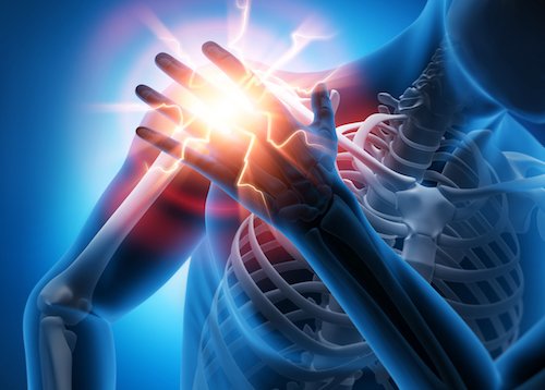
Complex Trauma Injuries
Treatment in Sharjah
Advanced Stabilization. Structural Reconstruction. A Real Path Back to Mobility.
When a single accident fractures more than one bone, tears through skin and muscle, or shatters a joint beyond simple repair, every hour matters. Dr. Ahmed Azmy provides specialized complex trauma injury care in Sharjah, Ajman and across the UAE, combining rapid emergency stabilization with advanced reconstructive surgery to protect limbs, restore function, and give patients the best possible chance at a full recovery.
If an injury is happening now or there are signs of shock, bleeding or poor circulation, seek emergency department care immediately.


RECONSTRUCTION & RECOVERY
Rapid emergency stabilization and vital-signs prioritization from the first moment of injury
Restoring complex joint surfaces, pelvic rings, and areas of significant bone loss
Preserving blood flow, tissue viability, and nerve function to protect the limb
A structured, long-term recovery plan built to regain independent mobility

EVERY STRUCTURE COUNTS.
Complex Trauma Injuries: More Than a Single Broken Bone
Complex trauma injuries are severe orthopedic injuries, usually from high-energy events such as car collisions, falls from height or heavy machinery accidents. They involve more than one broken bone: several fractures at once, shattered joints, extensive muscle and skin damage, or harm to nerves and blood vessels. Unlike a simple fracture, several structures fail together, so treatment has to cover all of them, often in stages rather than one operation. The first few hours after injury, "The Golden Window", matter most. Fast assessment and stabilization by an experienced surgeon often decide between a good recovery and lifelong disability.
What Makes Trauma Complex?
Complex trauma injuries describe a category of high-energy musculoskeletal damage in which the skeleton, joints, and surrounding soft tissues are affected together rather than in isolation. Unlike a straightforward fracture, these injuries typically involve more than one anatomical system, more than one break, or damage severe enough to threaten the survival of the limb itself. Understanding the anatomical scope of these injuries helps explain why they require a fundamentally different, and far more urgent, treatment approach.
 Anatomical & Structural Scope
Pelvic and Acetabular Disruptions
The pelvic ring and the acetabulum (the hip socket) can fracture or become unstable under high-energy impact, threatening both weight-bearing ability and the major blood vessels that run through the pelvis.
Severe Intra-Articular Shattering
Fractures that extend deep into a joint, the knee, ankle, shoulder, or elbow, break the smooth cartilage surface into multiple fragments, putting long-term joint function directly at risk.
Open & Compound High-Grade Fractures
Some fractures are severe enough that the broken bone breaks through the muscle and skin, exposing the fracture site to the outside environment and a high risk of contamination and infection.
Polytrauma
It is common in high-energy accidents for more than one limb, or more than one bone, to be fractured at the same time, requiring coordinated treatment planning across the entire skeleton, not just one injury site.
Periprosthetic Fractures
Patients who already have an artificial hip or knee replacement can sustain a new fracture around the implant, which requires specialized fixation techniques that respect the existing hardware.
Soft-Tissue Compromise
Complex trauma rarely affects bone alone. Surrounding muscle can lose its blood supply (necrosis), tendons can tear, and the vascular supply to the limb can be directly threatened, all of which must be assessed and treated alongside the fracture itself.
Types of Complex Orthopedic Trauma
Complex orthopedic trauma is classified by injury energy, fracture stability, and soft-tissue damage (skin, muscle, and blood vessels). This determines how urgently treatment is needed and how it is sequenced.
| Injury Pattern | Affected Anatomy | Clinical Features & Functional Risk |
|---|---|---|
| Pelvic ring fractures & dislocations | Pelvis, sacroiliac joints | Life-threatening internal bleeding risk; instability affects standing, walking, and sitting |
| Acetabular fractures (hip socket) | Hip joint, acetabulum | High risk of post-traumatic hip arthritis if the joint surface is not precisely restored |
| Comminuted tibial plateau / pilon fractures | Knee and ankle joint surfaces | Multiple bone fragments at the joint line; high risk of stiffness and early arthritis |
| High-grade open extremity fractures (Gustilo-Anderson grades) | Any long bone, with skin/muscle breach | Elevated infection risk; may require staged soft-tissue coverage |
| Multi-ligament knee dislocation with fracture | Knee ligaments plus adjacent bone | Risk to the popliteal artery behind the knee; joint instability |
| Segmental long bone fractures | A single bone broken in two or more separate levels | Complex fixation planning; higher risk of delayed healing |
| Severe foot & calcaneal crush injuries | Heel bone (calcaneus), midfoot | Long-term impact on walking pattern and weight-bearing comfort |
| Floating joint injuries (e.g., floating knee or elbow) | Fractures above and below a single joint | The joint is left "floating" and unsupported, requiring fixation at both ends |
| Nonunion or infected fracture pseudarthrosis | Any previously fractured bone that has failed to heal | Chronic complex trauma requiring revision surgery, often with infection control |
Pelvic & Major Joint Disruptions
Injuries to the pelvis, hip socket, and major weight-bearing joints, where precision reconstruction is essential to preserve long-term mobility.
High-Velocity Limb Crush & Open Wounds
Injuries where bone has broken through skin, or where a limb has been crushed, demanding urgent infection control and tissue viability assessment.
Multi-Site Skeletal Polytrauma
Cases involving multiple fracture sites across different bones or limbs, requiring coordinated, whole-body treatment sequencing.
Causes & High-Impact Mechanisms
Complex trauma injuries almost always result from a major transfer of energy, far greater than a simple slip or twist. Knowing the mechanism helps the surgical team anticipate hidden damage, since high-energy events often cause more internal injury than is visible.
Understanding the force of injury helps uncover damage that is not immediately visible.
Motor Vehicle & High-Speed Highway Collisions
Road traffic collisions remain one of the leading causes of complex orthopedic trauma, particularly at highway speeds where deceleration forces are extreme.
Motorcycle & Pedestrian Accidents
Riders and pedestrians have far less protection than vehicle occupants, making them especially vulnerable to multi-bone fractures and open wounds.
Falls from Substantial Heights
Falls from scaffolding, rooftops, or other elevated structures, common in industrial and construction settings, transmit enormous force through the pelvis, spine, and lower limbs.
Heavy Machinery & Crush Incidents
Industrial and agricultural machinery accidents can crush a limb between moving parts, causing combined bone, soft-tissue, and vascular injury.
Severe Sports Impacts & High-Speed Athletic Collisions
High-speed sports such as motorsport, equestrian activities, and contact sports can generate collision forces comparable to road traffic accidents.
Fragility-Induced Complex Fractures
In patients with significant osteoporosis (weakened, fragile bone), even a moderate-energy fall can result in a multi-fragment fracture pattern that behaves like a high-energy injury.
Complex Trauma Symptoms
The symptoms of complex trauma injuries are usually impossible to miss, but recognizing which signs point to a limb-threatening emergency, rather than "just" a bad break, is critical to seeking the right level of care quickly.
Severe Unrelenting Pain & Shock
Pain that is disproportionate to what would be expected, often accompanied by signs of systemic shock such as paleness, sweating, rapid heartbeat, or dizziness.
Obvious Gross Deformity & Limb Shortening
The limb may appear visibly bent at an unnatural angle, rotated, or shortened compared to the uninjured side.
Extensive Tissue Bruising, Blistering, and Massive Swelling
Rapid, significant swelling and skin changes reflect the scale of soft-tissue damage occurring beneath the surface.
Weak or Missing Pulse & Cold Hand or Foot
A pulse that is weak, absent, or a hand/foot that feels unusually cold can indicate the blood supply to the limb has been disrupted, a true surgical emergency.
Loss of Sensation, Drop Foot, or Severe Nerve Numbness
Numbness, tingling, or an inability to lift the foot or fingers can point to nerve injury alongside the fracture.
Inability to Move or Bear Any Weight on the Affected Area
A complete inability to use the limb, distinct from simple pain-limited movement, is a strong indicator of significant structural instability.
Complex Trauma Diagnosis & Physical Testing
Diagnosis combines a hands-on "complex trauma test" with advanced imaging, so the surgical team sees the full extent of the damage before planning treatment.
Primary ATLS (Advanced Trauma Life Support) Survey & Limb Viability Audit
A structured, whole-body assessment to identify and prioritize any immediately life- or limb-threatening problems.
Neurovascular Examination
Careful checking of pulses, capillary refill time (how quickly color returns to the skin after light pressure), and mapping of any nerve-related sensation or movement loss.
Soft-Tissue Envelope & Compartment Pressure Testing
Physical assessment of muscle tightness and, where needed, direct measurement of pressure inside muscle compartments to rule out compartment syndrome.
Stability Stress Assessments
Gentle, controlled physical tests to check pelvic stability and whether a joint is subluxating (partially dislocating) under stress.
Whole-Body Trauma CT Scans & 3D Reconstructions
Essential for mapping every fracture fragment in three dimensions and planning the reconstruction in advance.
High-Resolution Digital X-Rays
A trauma series that images the joint above and below the injured zone, since injuries frequently extend further than they first appear.
CT Angiography
A specialized scan that highlights the blood vessels, used to check for arterial tears or blockages following the injury.
MRI Evaluation
Used to assess soft-tissue structures, ligament avulsions (tears where a ligament pulls away from bone), and joint damage that may not be visible on X-ray or CT.

| Diagnostic Test | Clinical Role in Complex Trauma |
|---|---|
| Primary ATLS Survey | Identifies immediate life- and limb-threatening priorities |
| Neurovascular Examination | Confirms blood flow and nerve function are intact |
| Compartment Pressure Testing | Rules out or confirms acute compartment syndrome |
| Pelvic & Joint Stability Stress Testing | Assesses structural instability requiring urgent stabilization |
| Whole-Body Trauma CT & 3D Reconstruction | Maps fracture fragments for precise surgical planning |
| Digital X-Ray (Trauma Series) | Confirms fracture location, pattern, and alignment |
| CT Angiography | Detects arterial injury or vascular compromise |
| MRI | Evaluates soft-tissue, ligament, and occult joint damage |
When Should You See an Orthopedic Trauma Specialist?
Emergency room care and specialist orthopedic trauma care serve two different, and both essential, roles. The emergency room's priority is immediate resuscitation and life-saving stabilization. An orthopedic trauma specialist takes over from there, focusing on the precise, staged reconstruction needed to restore long-term limb function. If any of the following apply, a dedicated evaluation with a trauma specialist is strongly advised.
If you have cold or pale fingers or toes, significant bleeding, an open wound, shock or rapidly worsening pain: go to emergency services immediately.
Red Flag Signs for Immediate Expert Trauma Referral
- Bone fragments that remain unaligned following an initial ER cast or splint
- An open skin wound with visible bone, or bone previously exposed to the air
- Worsening nerve tingling, cold fingers or toes, or pale skin developing after initial treatment
- Bone that has not healed after several months (a possible nonunion or malunion)
- The need for secondary reconstruction after an initial emergency external fixator
Complex Trauma Treatment & Management
Complex fracture management is rarely a single operation, it is a carefully sequenced, staged process designed first to save the limb and the patient, and only then to rebuild it for the long term. Dr. Azmy's approach follows the internationally recognized principle of damage control first, definitive reconstruction second.
Emergency Stabilization
The immediate priority is controlling the situation, not perfecting it. This includes spanning external fixators (temporary frames that hold bones in position across a joint), thorough wound debridement (removing contaminated or dead tissue), infection control, and fasciotomies to release pressure when compartment syndrome is present.
Definitive Internal Fixation
Once the patient and the soft tissues are stable, definitive surgery is performed. This may involve ORIF (open reduction and internal fixation) with anatomical locking plates, retrograde or antegrade intramedullary nailing (rods placed inside the bone canal), bone grafting to fill gaps from bone loss, or 3D-guided osteosynthesis for the most complex fracture patterns.
Soft-Tissue Coverage & Microvascular Collaboration
Where skin and muscle loss is significant, reconstructive techniques, often performed in close collaboration with microvascular and plastic surgery colleagues, restore healthy tissue coverage over the repaired bone.
Joint Reconstruction or Primary Arthroplasty
When a joint surface has been too severely shattered to be salvaged through fixation alone, joint reconstruction or a primary joint replacement may offer the best path to a functional, pain-free joint.
| Treatment Phase | Surgical Strategy | Primary Objective |
|---|---|---|
| Stage 1: Emergency Stabilization | External fixation, debridement, fasciotomy | Save life and limb; control contamination |
| Stage 2: Definitive Internal Fixation | Locking plates, IM nailing, bone grafting | Restore bone alignment and stability |
| Stage 3: Soft-Tissue Coverage | Microvascular and plastic surgery collaboration | Protect repaired bone with healthy tissue |
| Stage 4: Joint Reconstruction / Arthroplasty | Joint reconstruction or replacement | Restore a functional, pain-free joint |
Advanced Fixation Technologies Used
Recovery Timeline and Long-Term Rehabilitation
Recovery from complex trauma injuries is a genuine journey, not a single milestone, and every patient's timeline will vary depending on the severity of the original injury. The table below offers a realistic general guide to what recovery typically looks like.
Hospital & Early Post-Operative
Initial stabilization complete; strict non-weight-bearing
Pain management, wound monitoring, early mobility of unaffected joints
Soft Tissue & Union Induction
Early bone healing (callus) begins; continued protection
Swelling gradually reduces; regular wound and X-ray review
Protected Mobility & Callus Formation
Partial weight-bearing may begin, guided by imaging
Increasing confidence in gentle movement, still supervised closely
Dynamic Loading & Progressive Independence
Progression toward full weight-bearing
Return of daily function; structured physiotherapy intensifies
Long-Term Remodeling & Functional Return
Bone remodeling continues; strength and endurance rebuild
Return to work, sport, or full independence, guided by ongoing review
Factors That Influence Healing in Polytrauma
Adequate blood supply is essential for bone and tissue healing
Significantly slows bone union and increases complication risk
Protein, vitamin D, and calcium intake directly support bone repair
Greater soft-tissue or bone loss generally extends the overall recovery timeline
Specialized Trauma Rehabilitation & Exercises
Rehabilitation after complex trauma is progressive by design, every stage builds the foundation for the next, and skipping ahead too quickly can undo months of careful surgical work. All exercise progression should be guided by a supervising physiotherapist in coordination with the surgical team.
Early Edema Control & Passive Bed Mobility
Gentle positioning and passive movement, guided by the care team, to reduce swelling and prevent deep vein thrombosis (DVT) and joint stiffness in the earliest recovery days.
Gentle Muscle-Tightening Exercises
Muscle-tightening exercises that don't move the joint itself, used to protect the surgical repair while still stimulating healthy blood flow to the area.
Active-Assisted Joint Mobilization
Carefully guided joint movement, supported by a therapist or equipment, to safely begin restoring range of motion once healing allows.
Balance & Walking Retraining
Hydrotherapy (exercise in water), parallel bar walking practice, and adaptive balance training to rebuild the body's sense of position and safe, confident walking.
Complications of Untreated or Inadequately Managed Trauma
Complex trauma injuries that are not treated promptly, or not managed by a team experienced in high-energy orthopedic trauma, carry a meaningfully higher risk of long-term complications.
Nonunion (Bone That Won't Heal )
The bone fails to heal together at all
Malunion (Bone Healed Crooked)
The bone heals, but in a crooked or misaligned position
Post-Traumatic Osteoarthritis ( Joint Wear After Injury)
Early breakdown of joint cartilage following joint-surface injury
Chronic Osteomyelitis (Long-Lasting Bone Infection )
A persistent, deep bone infection that can be difficult to fully clear
Volkmann's Ischemia (Lasting Muscle Damage from Pressure )
Long-term muscle damage and contracture from unresolved compartment pressure
Heterotopic Ossification (Extra Bone Growth in Muscle )
Abnormal bone formation within muscle or soft tissue around the injury
Complex Regional Pain Syndrome (CRPS)
A chronic pain condition that can develop after significant limb trauma
Limb Length Discrepancy & Pelvic Tilt
A lasting difference in limb length or pelvic alignment affecting posture and gait
Why Choose Dr. Ahmed Azmy in Sharjah?
When an injury is complex, experience matters. Dr. Ahmed Azmy combines 15+ years of trauma surgery, German fellowship training, and a limb salvage approach to give patients the best chance of full recovery.
- German fellowship training, with particular expertise in joint reconstruction and minimally invasive trauma fixation
- A dedicated limb salvage philosophy, prioritizing preservation of the patient's own limb wherever safely possible
- Comprehensive post-trauma rehabilitation pathways, coordinated from surgery through full functional recovery
- Close coordination with leading surgical facilities across Sharjah and the UAE
The Patient-Centred Trauma Pathway
Emergency Intake Review
A thorough review of the injury, prior imaging, and emergency treatment already received
Clear Family Communication
Honest, understandable explanations of the injury, the plan, and realistic expectations
Precision Surgery
Staged, meticulously planned surgical care tailored to the individual injury pattern
Dedicated Functional Recovery
Ongoing rehabilitation support focused on returning patients to independent living
Patient Testimonials & Recovery Stories
These narratives were supplied in the page draft and require patient consent and source verification before they are published as real testimonials.
Publishing note: Do not present these draft narratives as independently verified patient reviews. Replace them with consented genuine testimonials, or omit this section.
Frequently Asked Questions
Clear answers on fracture complexity, fixation, diagnosis, treatment and long-term recovery.
What exactly qualifies as a complex trauma injury?
A complex trauma injury generally involves more than one fracture, a joint surface broken into multiple fragments, an open wound exposing the bone, or damage to the nerves and blood vessels around the injury — as opposed to a single, simple, isolated break.
What is an external fixator, and why is it used before permanent surgery?
An external fixator is a temporary metal frame, attached to the bone through the skin, that holds a severely injured limb in a stable position. It is used first because it stabilizes the injury quickly and safely while the soft tissues recover enough to tolerate more definitive internal surgery.
How long does recovery take after complex orthopedic trauma?
Recovery timelines vary widely depending on injury severity, but patients typically progress through hospital care, protected mobility, and progressive weight-bearing over several months, with full bone remodeling and functional return continuing for up to 12–18 months or more in severe cases.
What is the difference between a simple fracture and complex trauma?
A simple fracture usually involves one clean break in one bone, often treated with a cast and predictable healing. Complex trauma involves multiple injuries occurring together, such as several fractures, joint damage, and soft-tissue or vascular injury, requiring staged surgical management.
What is an orthopedic complex trauma test or evaluation?
This refers to the physical and diagnostic examination battery used to assess a trauma patient, including neurovascular checks, compartment pressure testing, joint stability stress tests, and imaging such as CT and MRI — used to map the full extent of the injury before treatment planning.
Can a shattered joint recover without undergoing total joint replacement?
In many cases, yes. If enough of the joint surface can be precisely reconstructed and fixed, function can often be preserved without replacement. However, in cases of very severe joint destruction, reconstruction or replacement may offer the more reliable long-term outcome.
What are the signs of infection or hardware failure after trauma surgery?
Warning signs include increasing pain rather than improving pain, redness, warmth, or drainage at the surgical site, fever, or new instability or movement at the fracture site. Any of these should prompt urgent evaluation.
When should I seek a second opinion for a fracture that is not healing properly?
If a fracture has not shown clear signs of healing on imaging within the expected timeframe for that injury, often around 3–6 months, or if pain and instability persist, it is reasonable and encouraged to seek a specialist second opinion promptly.
Restore Mobility and Rebuild Quality of Life
A high-impact injury is frightening, for you and your family. With Dr. Ahmed Azmy's team and the right treatment sequence, a real return to independence and mobility is achievable. Take the first step today.
Fast, accurate stabilization from the moment of injury
A dedicated commitment to saving and restoring the patient's own limb
Precision surgical techniques matched to each unique injury pattern
Structured, supervised recovery built around long-term function