Lower Limb Fractures Treatment in Sharjah
Specialist diagnosis, bone realignment and mobility-focused rehabilitation for hip, thigh, knee, leg, ankle and foot injuries.
A broken limb in the lower body does more than cause pain, it can take away your ability to stand, walk and stay independent overnight. Dr. Ahmed Azmy provides specialist assessment, weight-bearing imaging review, accurate bone realignment, surgical fixation when required, and a structured path back to safe mobility for patients in Sharjah, Ajman and across the UAE.

What Are Lower Limb Fractures?
A lower limb fracture is a partial or complete break in one or more of the weight-bearing bones extending from the hip to the toes. Because these bones support your entire body weight, even a single fracture can affect standing, walking and everyday independence until it is properly diagnosed and treated.
The injury may involve a simple hairline crack, a fully displaced bone, several bone fragments, or a fracture that extends into a nearby joint. Lower limb fractures can also damage the surrounding ligaments, tendons, muscles, nerves and blood vessels, which is why treatment must always consider the whole limb rather than the broken bone in isolation.
A fracture may be closed, meaning the skin remains intact, or open, meaning the broken bone communicates with a wound through the skin. Open lower limb fractures are orthopedic emergencies because of the high risk of contamination and infection, and require immediate hospital-based treatment.

Hip & Pelvis
The ball-and-socket joint and surrounding pelvic bones that connect the leg to the spine and support the body's weight.
Femur (Thigh Bone)
The longest and strongest bone in the body, running from the hip to the knee.
Patella & Knee Joint
The kneecap and the joint surfaces of the femur and tibia that allow the knee to bend and straighten.
Tibia & Fibula (Shin/Calf)
The two bones of the lower leg, with the tibia bearing most of the body's weight.
Ankle (Malleoli & Syndesmosis)
The joint formed by the tibia, fibula and talus, stabilised by the malleoli and syndesmosis ligaments.
Foot & Toes
Including the calcaneus (heel bone), talus, metatarsals and toe bones that absorb impact with every step.
Because the lower limb is responsible for standing, walking and weight-bearing, early orthopedic assessment matters far more here than in many other injuries. A missed or delayed diagnosis can allow abnormal weight distribution, worsening displacement or circulation problems to develop before treatment even begins.
Types of Lower Limb Fractures
Lower limb fractures are classified according to the bone involved, the exact location of the break, whether the fragments remain aligned, and whether the fracture extends into a joint surface. The table below summarises the most common lower limb fractures in patient-friendly language.

Hip & Thigh Fractures
Femoral neck, intertrochanteric, shaft and distal femur injuries affecting standing and transfer safety.

Knee & Lower Leg Fractures
Patella, tibial plateau, tibial shaft and fibula fractures with major mobility consequences.

Ankle & Foot Fractures
Ankle, calcaneus, talus and metatarsal fractures that directly affect gait and balance.
| Fracture Type | Location | Clinical Description |
|---|---|---|
| Hip fracture (Femoral neck & intertrochanteric) | Top of the thigh bone at the hip | Usually follows a fall, especially in older adults with reduced bone density. Causes severe groin or hip pain and an inability to stand or walk. |
| Femoral shaft fracture | Middle of the thigh bone | Often the result of high-energy trauma. Produces severe pain, deformity and significant swelling of the thigh. |
| Distal femur fracture | Lower thigh bone near the knee | A complex fracture close to the knee joint that can make bending the knee extremely painful or impossible. |
| Patellar fracture | Kneecap | Often follows a direct fall onto the knee or a sudden forceful contraction of the thigh muscle. Straightening the knee becomes difficult. |
| Tibial plateau fracture (Knee joint) | Top of the shin bone at the knee | A joint-surface fracture that can affect long-term knee stability and function if not carefully realigned. |
| Tibial shaft fracture | Middle of the shin bone | A common lower leg fracture, frequently from direct impact or twisting injuries, that can affect weight-bearing significantly. |
| Fibula fracture | Outer bone of the lower leg | May occur alone or alongside a tibial fracture. Often follows a twisting or direct-impact injury near the ankle. |
| Bimalleolar / Trimalleolar ankle fracture | Ankle joint | Involves two or three bony prominences around the ankle and usually causes marked swelling, deformity and instability. |
| Calcaneus (Heel bone) fracture | Heel | Typically follows a fall from height landing directly on the heel. Causes severe heel pain and difficulty bearing weight. |
| Talus fracture | Ankle bone beneath the tibia | A less common but serious fracture that can threaten the blood supply to part of the bone if not managed promptly. |
| Metatarsal fractures (incl. Jones & stress fractures) | Midfoot bones | May follow a direct blow, twisting injury or repetitive overload. Causes localised pain and difficulty walking. |
| Lower limb fracture-dislocation | Hip, knee or ankle | The bone is broken and the joint surfaces have also lost their normal alignment. Requires urgent reduction. |
| Open lower limb fracture | Any lower-limb bone | The fracture communicates with a skin wound. This is an orthopedic emergency due to contamination and infection risk. |
Priority Injury Group 01
Hip & Thigh Fractures- Femoral neck fracture
- Intertrochanteric fracture
- Femoral shaft fracture
- Distal femur fracture
Priority Injury Group 02
Knee & Lower Leg Fractures- Patellar fracture
- Tibial plateau fracture
- Tibial shaft fracture
- Fibula fracture
Priority Injury Group 03
Ankle & Foot Fractures- Bimalleolar / trimalleolar ankle fracture
- Calcaneus fracture
- Talus fracture
- Metatarsal fractures
Lower Limb Fracture Symptoms
Symptoms usually begin immediately, though stress fractures may develop more gradually. Pain severity alone doesn't indicate stability, every suspected fracture needs proper assessment.

Inability to Bear Weight
- Cannot stand, walk or put pressure on the leg
- Instinctively shifts weight to the uninjured side
- Leg feels unstable or gives way
Severe Pain & Tenderness
- Sharp, throbbing pain over the fracture
- Pain aggravated by movement or weight-bearing
- Tenderness on direct touch over the bone
Rapid Swelling & Bruising
- Swelling develops quickly after injury
- Bruising spreading into the ankle or foot
- Tightness in the skin as swelling increases
Visible Deformity
- Abnormal angle of the leg, knee or ankle
- Leg shortening vs. the uninjured side
- Rotated or abnormally positioned foot
Crepitus & Restricted Motion
- A grinding sensation when the leg is moved
- A locked or blocked joint
- Marked resistance to bending knee/ankle
Nerve or Blood Supply Signs
- Tingling or numbness in the foot/toes
- Cold or pale toes vs. the other foot
- A weak or absent pulse in the foot
New numbness, tingling, pale or cold toes, a weak pulse, or increasing tightness in the leg require urgent assessment. These signs can indicate impaired circulation, nerve compression or acute compartment syndrome, all of which need immediate medical attention rather than observation at home.
Causes of Broken Lower Limbs
Lower limb fractures can result from a single high-energy traumatic event or, less commonly, from repetitive loading over time. Understanding the mechanism of injury helps predict associated damage and guides the choice of imaging and treatment.
Mechanism matters
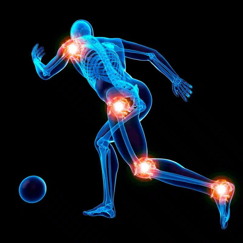
Trauma energy, direction of force and the loading pattern all help predict the fracture pattern and the associated soft-tissue risk.
High-Velocity Road Traffic Collisions
Car and motorcycle trauma can transmit enormous force, frequently producing displaced, open or multiple fractures.
Falls from Height & Direct Trauma
Landing directly on the feet or knees can fracture the heel, ankle, tibia or femur, with possible associated pelvic injury.
Sports & Twisting Injuries
Football tackles, running pivots and skiing falls can twist or strike the lower leg, ankle or knee.
Simple Slips & Trips in Older Adults
Reduced bone density from osteoporosis means a low-energy slip can be enough to fracture the hip or lower leg.
Repetitive Stress & Overuse
Runners and athletes can develop stress fractures in the tibia or metatarsals from repeated loading.
Stable vs. Unstable Lower Limb Fractures
Whether a lower limb fracture is stable or unstable determines whether a patient can be safely treated in a cast or walking boot, or whether surgical fixation is needed to protect alignment, weight-bearing capacity and long-term joint function.
| Clinical Factor | Stable Fracture | Unstable Fracture |
|---|---|---|
| Bone alignment | Fragments remain in an acceptable position | Fragments are displaced, angulated, shortened or rotated |
| Weight-bearing capability | Can often tolerate protected or partial weight-bearing | Weight-bearing is unsafe until stabilised |
| Joint surface involvement | Joint surface remains smooth and congruent | Fracture enters and disrupts the joint surface |
| Soft tissue condition | Skin and surrounding tissues remain intact | Open wound, threatened skin or major soft-tissue injury |
| Blood supply & nerve status | Normal foot pulses, sensation and circulation | Reduced pulse, numbness or impaired circulation |
| Typical management | Cast, walking boot, brace and monitored rehab | Surgical fixation or reduction is usually required |
Stable Fracture Features
- Acceptable alignment
- Minimal displacement
- No neurovascular compromise
- Can be managed in a cast or boot
- Confirmed by follow-up imaging
Unstable Fracture Features
- Significant displacement
- Rotation or shortening
- Joint-surface involvement
- Open wound or threatened skin
- Impaired circulation or nerve function
Lower Limb Fracture Diagnosis
A thorough diagnosis identifies far more than the presence of a fracture. It establishes the fracture pattern, fragment position, joint involvement, stability, and whether the surrounding nerves, blood vessels or soft tissues have been affected.
It is not just about confirming the fracture — it is about understanding stability, alignment, joint involvement and tissue safety.
Patient History & Injury Mechanism
How the injury occurred helps predict the likely fracture pattern and associated injuries.
Physical Examination
Deformity, skin tenting, swelling and localised tenderness identify the injured region and urgency.
Neurovascular & Pulse Assessment
Checking the dorsalis pedis pulse and toe sensation detects compromise to blood flow or nerves.
Adjacent Joint & Soft Tissue Check
Knee and ankle stability plus compartment firmness identify nearby injuries or early compartment syndrome.
Weight-Bearing / Standard X-Rays
The primary investigation for confirming most fractures and assessing alignment and joint involvement.
CT Scan
Essential for complex intra-articular fractures like tibial plateau, calcaneus and pilon fractures.
MRI Scan
Used for occult stress fractures or suspected combined ligament/cartilage damage.
| Imaging Test | Best Used For |
|---|---|
| Weight-Bearing / Standard X-Ray | Confirming most fractures and assessing alignment, displacement and joint involvement. |
| CT Scan | Defining complex, comminuted and intra-articular fractures (tibial plateau, calcaneus, pilon) and surgical planning. |
| MRI Scan | Identifying occult stress fractures and associated ligament, cartilage or soft-tissue injury. |
When to Seek Immediate Medical Attention
Some lower limb fracture symptoms are true emergencies. Knowing the warning signs can protect circulation, nerves and the limb itself.
Emergency Warning — Obtain emergency medical care immediately if:
- Open fractures — bone piercing the skin or visible through a wound
- Acute Compartment Syndrome — extreme, unrelenting leg pain, stone-hard muscles, pain on passive toe stretch
- Loss of circulation — cold, pale or blue toes, or an absent pulse
- Severe deformity, limb shortening or a dislocated joint
Immediate First-Aid Guidance
- Immobilise the leg in the position found
- Do not attempt to bear weight on the injured leg
- Do not push exposed bone back through the skin
- Apply covered ice, never directly onto the skin
- Elevate the leg if it is safe to do so
- Avoid eating or drinking if emergency surgery may be required
Treatment Options for Lower Limb Fractures
Treatment aims to restore alignment, protect nerves and circulation, and return the patient to safe mobility. The right approach depends on fracture pattern and stability, not pain intensity alone.
| Treatment Consideration | Conservative Non-Surgical Management | Advanced Surgical Management |
|---|---|---|
| Most appropriate for | Stable, non-displaced or acceptably aligned fractures | Displaced, unstable, open, intra-articular or fracture-dislocation injuries |
| Alignment | Satisfactory and maintainable without internal fixation | Requires reduction and mechanical stabilisation |
| Methods | Cast, walking boot, brace or closed reduction | Plates, screws, intramedullary nails or external fixation |
| Weight-bearing | Progressed according to fracture stability | Introduced according to fixation security and soft-tissue healing |
| Monitoring | Clinical review and repeat X-rays when required | Wound review, imaging and postoperative follow-up |
| Primary objective | Maintain alignment while natural bone healing occurs | Restore alignment and create stable fixation for healing and rehab |
Conservative Non-Surgical Management
- Rigid casts for tibial, ankle and selected foot fractures
- Walking boots (CAM boots) for controlled protected weight-bearing
- Braces for selected knee and lower leg injuries
- Crutches or walkers for non-/partial weight-bearing protocols
- Serial follow-up X-rays to confirm alignment during healing
Advanced Surgical Interventions
- ORIF — anatomical plates & screws
- Intramedullary Nailing — femur and tibia fractures
- External Fixation — severe soft-tissue compromise, open fractures, polytrauma
- MIPO — minimally invasive percutaneous osteosynthesis
- Joint Reconstruction or Arthroplasty — severe geriatric hip fractures
When Surgery is Indicated
- The bone is significantly displaced, angulated, rotated or shortened
- The fracture enters and disrupts a weight-bearing joint surface
- The fracture is unstable
- Closed reduction does not restore or maintain acceptable alignment
- The fracture is open or contaminated
- A nerve or blood vessel is compromised
- The injury is associated with a dislocation or major ligament injury
- Multiple fractures affect the same limb
- Early weight-bearing and mobility would otherwise be significantly delayed
Get a personalised lower limb fracture treatment plan in Sharjah.
Recovery Timeline and Functional Phases
Because the lower limb bears full body weight, recovery is governed by how weight-bearing is progressed. The timeline below is a general guide, actual recovery varies by bone, pattern and patient.
Emergency protection
Immobilisation, imaging, pain control, neurovascular checks
Significant pain, swelling and inability to bear weight
Early stabilisation
Cast/wound review; strict non-weight-bearing where indicated
Pain begins to settle; leg remains protected
Early bone healing
Non-weight-bearing or partial weight-bearing with crutches
Bruising resolves; stiffness may develop
Progressive weight bearing
Gradual increase to fuller weight-bearing under guidance
Confidence in standing improves
Full weight bearing & gait retraining
Structured gait training and progressive strengthening
Daily tasks become easier; some residual weakness
Advanced strength & sports return
Advanced strengthening, balance work, sport-specific progression
Many resume broader activity after clearance
Four functional recovery phases
Protection, Edema Control & Safe Transfers
Immobilisation, swelling reduction, safe non-weight-bearing transfers.
Range of Motion & Partial Weight-Bearing
Controlled joint movement plus partial weight-bearing when allowed.
Full Weight-Bearing & Muscle Strengthening
Progressive loading with thigh, calf and hip strengthening.
Gait Normalization & Return to Activity/Sport
Retraining normal walking, balance, endurance, and graded return to sport.
Lower Limb Rehabilitation Exercises
The following exercise categories should be performed strictly under specialist guidance, as the appropriate stage and intensity depend on the specific fracture and its healing progress.
Early Circulation & Mobility
- Ankle pumps
- Gentle quad sets
- Straight leg raises when cleared
Intermediate Strength
- Seated heel-toe raises
- Supported mini-squats
- Resistance band work
Advanced Balance & Gait Retraining
- Proprioception board work
- Stationary cycling
- Step-ups
Exercises should never be forced or advanced before radiographic union is confirmed. Progressing too quickly can disturb healing bone and increase the risk of displacement or refracture.
Complications of Untreated or Improperly Managed Fractures
A lower limb fracture that is not diagnosed, reduced or monitored appropriately can affect walking ability for years. Early treatment and structured follow-up help reduce avoidable deformity, joint damage, circulation problems and chronic pain.
Malunion
Bone heals in an abnormal position, leading to limb length discrepancy, limping and accelerated joint wear.
Nonunion or Delayed Union
Slow or failed healing causing ongoing pain, instability and possible hardware failure.
Acute Compartment Syndrome
Rising pressure in the leg's muscle compartments can permanently damage muscles and nerves if untreated.
Post-Traumatic Osteoarthritis
Fractures into the hip, knee or ankle joint surface increase long-term risk of joint wear.
DVT & Pulmonary Embolism
Reduced mobility increases the risk of leg blood clots, which can potentially travel to the lungs.
Joint Stiffness & Muscle Atrophy
Prolonged immobilisation can reduce range of motion and cause thigh or calf muscle wasting.
Avascular Necrosis (AVN)
Certain fractures (femoral neck, talus) can interrupt blood supply, leading to bone collapse.
Chronic Regional Pain Syndrome (CRPS)
Disproportionate, persistent pain, swelling and temperature/colour changes requiring early management.
Why Choose Dr. Ahmed Azmy in Sharjah?
Lower limb trauma demands precise decision-making. Dr. Ahmed Azmy focuses on more than bone healing, restoring alignment, protecting circulation and nerves, and rebuilding confident, independent walking.
years of orthopedic and trauma experience
patients treated
procedures performed
fellowship-trained expertise
minimally invasive techniques that preserve blood supply and soft tissues
Key highlights: Minimally invasive techniques that preserve blood supply and soft tissues · Comprehensive rehabilitation and weight-bearing protocols.
Accurate Evaluation
Assessment considers injury mechanism, fracture pattern, soft-tissue condition, circulation and functional priorities.
Clear, Non-Jargon Explanations
Direct explanation of stability, joint involvement, treatment options and warning signs in plain language.
Personalised Surgical/Conservative Plan
Treatment matched to the specific fracture pattern, bone quality and daily mobility needs.
Structured Return-to-Walking Guide
A clear, staged rehabilitation plan that safely progresses weight-bearing, strength and gait.
Specialist lower limb fracture assessment for patients from Sharjah, Ajman and the UAE.
Patient Testimonials & Success Stories
Real patient journeys show how the right diagnosis and treatment plan can restore mobility, whether the case calls for surgery or conservative care.
Femoral Shaft Fixation After a Traffic Accident
“My thigh bone was fractured in a car accident, and I couldn't stand or move without severe pain. Dr. Ahmed Azmy explained the fixation clearly and guided me through a structured rehabilitation programme. I gradually regained my strength and I'm walking confidently again.”
Bimalleolar Ankle Fracture Recovery
“I fractured my ankle badly after a fall and was worried I might never walk normally again. The treatment plan and follow-up care were thorough, and my progressive weight-bearing programme helped me return to daily activities with confidence.”
Conservative Management of a Stress Fracture
“As a runner, I developed persistent shin pain that turned out to be a stress fracture. Dr. Ahmed Azmy managed it without surgery, with a clear plan for rest, protected loading and a gradual return to running.”
Frequently Asked Questions
1. How long does a broken leg or lower limb fracture take to heal?
Healing time varies according to the bone involved, fracture stability, patient age, bone quality and treatment method. Simple lower leg or ankle fractures may take around 6 to 12 weeks to heal sufficiently for full weight-bearing, while hip, femur or complex joint fractures can take several months. Your treating specialist will confirm progress with follow-up imaging rather than a fixed timeline alone.
2. How soon can I put weight on my leg after a lower limb fracture?
This depends entirely on the fracture pattern and stability. Some stable fractures allow protected partial weight-bearing within days, while others require several weeks of non-weight-bearing to protect alignment and fixation. Weight-bearing should only be increased under the guidance of your treating specialist.
3. When is surgery required for a lower limb fracture?
Surgery is more likely when the fracture is displaced, unstable, open, comminuted, extends into a joint surface, or cannot maintain acceptable alignment in a cast. It may also be recommended when circulation or nerve function is affected, or when early, safe weight-bearing could otherwise be significantly delayed.
4. What is the difference between a sprain and a hairline fracture in the ankle or foot?
Both can cause pain, swelling and difficulty walking, so symptoms alone are not reliable. A sprain affects the ligaments supporting the joint, while a hairline fracture is a crack in the bone itself and may not always be visible on an initial X-ray. Persistent or worsening pain should be assessed by a specialist.
5. Why are blood thinners sometimes prescribed after a lower limb fracture?
Reduced mobility increases the risk of blood clots forming in the leg veins (deep vein thrombosis), which can potentially travel to the lungs. Blood thinners may be prescribed for selected patients, particularly after surgery or prolonged immobilisation, to reduce this risk.
6. Can I drive with a lower limb cast or boot?
Driving is not safe while a cast, boot or brace restricts your ability to control the pedals or react quickly in an emergency, whichever leg is affected. Clearance to drive should be confirmed with your treating specialist based on your specific fracture and recovery stage.
7. How do I know if my leg cast or splint is too tight?
Warning signs include increasing pain, numbness or tingling in the toes, toes that appear pale, blue or unusually cold, and difficulty moving the toes. These symptoms should never be observed at home — contact your treating team promptly or seek emergency care if severe.
8. When can I return to sports or high-impact running after a lower limb fracture?
Return to sport requires more than the absence of pain. The fracture should show confirmed radiographic healing, and you should demonstrate adequate strength, balance, range of motion and confidence in weight-bearing before progressing to high-impact activity.
Injured your hip, thigh, knee, lower leg, ankle or foot?
Get expert evaluation and a personalised fracture treatment plan from Dr. Ahmed Azmy.